Figures & data
Table 1. Obstetric characteristics of the HELLP syndrome and control group.
Table 2. Hematological and biochemical parameters in both groups.
Table 3. Morphometric parameters in both groups.
Table 4. Morphometric graphics.
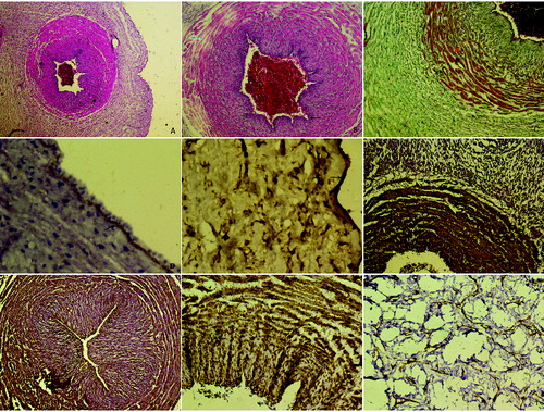
Figure 1. A: Control group, appearance normally of umbilical cord, H–E stain Bar 50 µm. B and C: HELLP group; degeneration of endothelial cell in the vessel wall (arrows), thickness in basal membrane, edema in the intermediate connective tissue between muscle layers (arrows), H–E stain Bar 100 µm, increase in collagen fibres between smooth muscle layers (arrow), Tichrom-Masson stain Bar 100 µm. D and E: Strong expression of MMP9 in basal membrane of vessel wall and amniotic epithelium (arrows). Also positive expression in Wharton's jelly. MMP9 immunohistochemistry staining Bar 100 µm. F and G: Positive expression in endothelial cells, basal membrane and fibroblast cells in HELLP group, CD44 immunohistochemistry staining Bar 50 µm. H and I: Positive reaction of actin protein in Wharton's jelly spindle-like fibroblast cells and smooth muscle layers. α-smooth muscle actin immunohistochemistry staining Bar 100 µm.