
Figures & data
Figure 1. Representative microscopic image showing (a) ‘no defect’ at 3 mm level, (b) ‘partial crack’ at 3 mm level, (c) ‘craze line’ at 6 mm level and (d) ‘fracture’ at 3 mm level.
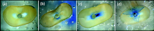
Figure 2. Distribution of number of dentinal defects at 3 mm root level (more than one defect per slice was possible).
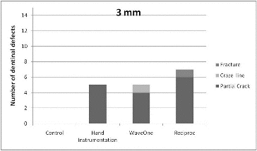
Figure 3. Distribution of number of dentinal defects at 6 mm root level (more than one defect per slice was possible).
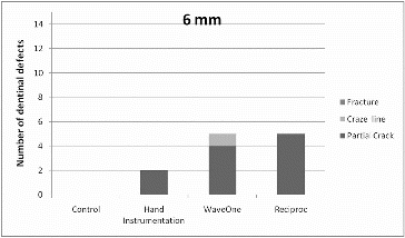
Figure 4. Distribution of number of dentinal defects at 9 mm root level (more than one defect per slice was possible).
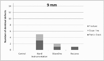
Table 1. Numbers of teeth presenting dentinal defects in different cross-sectional slices.