
Figures & data
Figure 1. The result of the EDS test on a histological plate for the determination of the metallic particles' nature.
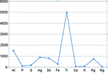
Figure 2. Mild inflammatory infiltration in the underlying connective tissue area (arrow).
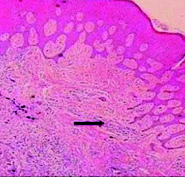
Figure 3. Severe infiltration of inflammatory cells in the connective tissue area (arrow).
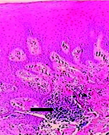
Figure 4. The presence of small metallic particles in the subepithelial connective tissue (upper arrow) and haemorrhagic areas in the connective tissue (lower arrow).
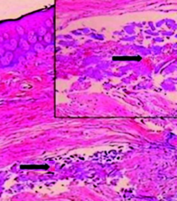
Table 1. Evaluation of the inflammatory statuses of the samples in terms of the metallic particles detected at the epithelium‒connective tissue junction.
Table 2. Evaluation of the inflammatory statuses of the samples in terms of the metallic particles detected at the connective tissue‒cover screw surface junction.
Table 3. Relationship between the presence of metallic particles in the samples and severity of inflammation at epithelium‒connective tissue junction, within the connective tissue and at connective tissue‒cover screw surface junction.