
Figures & data
Figure 1. Cohort of patients distributed into two subgroups according to age and gender.
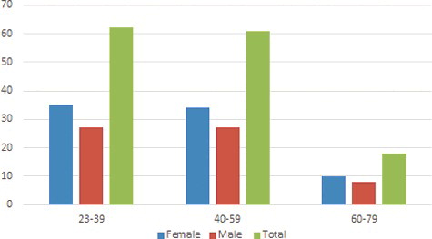
Figure 2. Three-dimensional CBCT (a), cross-sectional (b), panoramic reconstructed images (c) showing the mandibular canal and anterior loop.
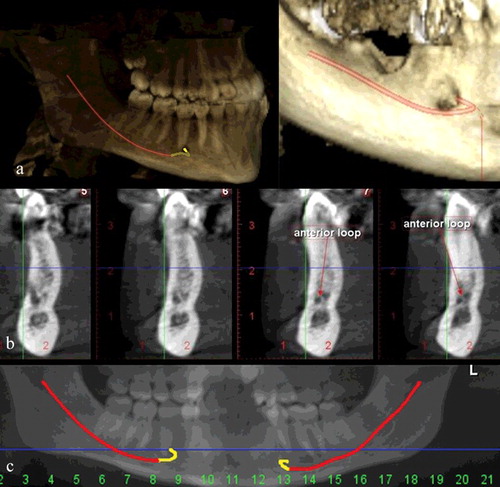
Figure 3. Cross-sectional (a) and sagittal (b) CBCT images showing the measurements for anterior loop morphology.
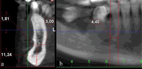
Figure 4. Volume of mandibular canal and anterior loop measured three-dimensionally (using 3D Invivo software).
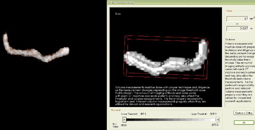
Table 1. Inter-observer consistency of measurements.
Table 2. Mean values of anterior loop measurements (mm) according to age groups and gender.
Table 3. Mean values (mm3) of anterior loop measurements according to age groups and gender.