Figures & data
Table 1. Comparison of CTx, TRACP 5b and ALP levels between the groups.
Table 2. Evaluation of serum biomarkers levels between non-ONJ and ONJ groups.
Table 3. Evaluation of clinical findings among the groups.
Figure 1. Clinical appearance of extraction sites: control group; complete mucosal healing, Group I; osteonecrosis with swelling and hyperaemia, Group II; osteonecrosis, Group III; complete mucosal healing.
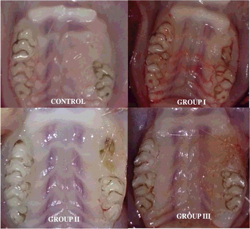
Figure 2. Histopathological view showing the normal and necrotic bone areas on the jaws: (A) control, normal bone cellularity, (B and C) Groups I and II, large necrotic areas (stars) bone, apoptotic osteoclasts with condensate nuclei (arrowhead), (D) Group III, small amounts of necrotic bone, control like histological features (HE, 100×).
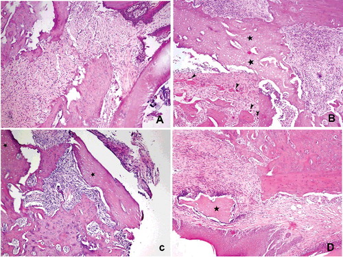
Figure 3. Evaluation of the ratio of necrotic bone area to the total bone area among the groups.
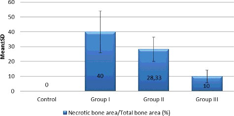
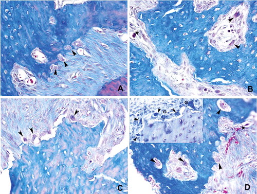
Figure 4. Histopathological view showing the osteoblastic activity: cuboidal and dense stained osteoblasts (arrow) reflected osteoid formation. Basophilic reversal lines (arrowhead) were considered to identify the bone remodelling (A) control, moderate osteoblastic activity, (B and C) Groups I and II, mild to moderate osteoblastic activities along with small amounts of reversal lines, (D) Group III, an increased osteoblastic activity (Toluidin blue 400x).
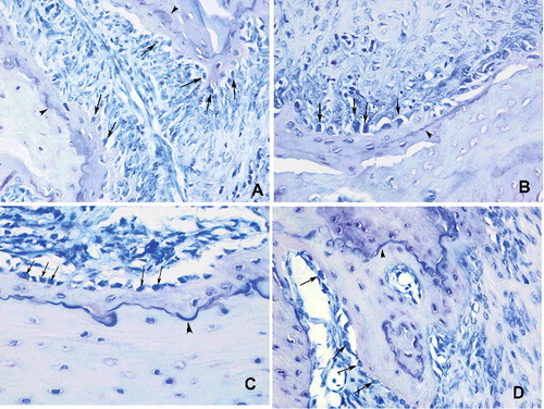
Figure 5. Histopathological view showing the severity of inflammation among the groups: (A) control, normal appearance, (B and C) Groups I and II, focal inflammation areas, neutrophils and foamy histiocytes surrounding the extraction sites, severe inflammation, (D) Group III, similar with control group, mild inflammation (PAS + HL 200×).
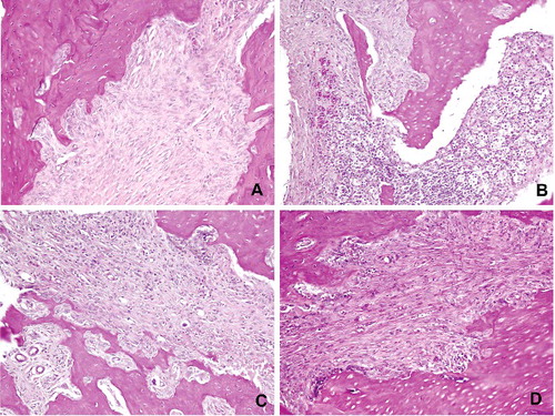
Table 4. Evaluation of severity of inflammation among the groups.
Table 5. Evaluation of osteoclast number among the groups.
Figure 6. Histopathological view showing the osteoclast density: (A) control, osteoclasts (arrowhead) were shown associated with bone surface in their active pole in the control group. (B) Detached (arrowhead) and apoptotic (star) osteoclasts were marked in Group I. (C and D) Osteoclast number with normal appearance was increased in Groups II and III. Giant type osteoclasts were characteristics of that groups (Mallory Triple 400x)