Figures & data
Table 1. Cav3.3 shRNA primers.
Figure 1. Band of the pYr-1.1-Cav3.3 shRNA plasmid digested with BsaI. Lane 1: pYr-Cav3.3 shRNA1; Lane 2: pYr-Cav3.3 shRNA2; Lane 3: pYr-Cav3.3 shRNA3; M: molecular size marker (Thermo Fisher Scientific Inc., Waltham, MA, USA).
Figure 2. Sequences (underlined) of pYr-1.1-Cav3.3 shRNA-1, pYr-1.1-Cav3.3 shRNA-2 and pYr-1.1-Cav3.3 shRNA-3.
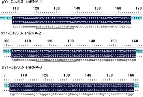
Figure 3. Production of recombinant adenovirus identified by PCR in 293 cells. Lane 1: pAd/PL-DEST vector; Lane 2: pAd-Cav3.3 shRNA; Lane 3: negative control.
Figure 4. pAd-Cav3.3 shRNA adenovirus infection efficiency in DRG cells: 24 h (A) and 48 h (B) after infection with pAd-Cav3.3 shRNA adenovirus and after amplification (C,D).
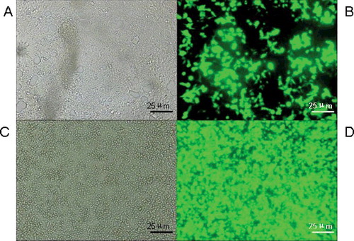
Figure 5. Expression of Cav3.3 mRNA.
Figure 6. Expression of Cav3.3 protein. Lane 1: expression of Cav3.3 protein in normal DRG cells; Lane 2: expression of Cav3.3 protein in empty DRG cells; Lane 3: expression of Cav3.3 protein in pAd-Cav3.3 shRNA adenovirus-infected DRG cells.