Figures & data
Figure 1. UPLC-DAD (A) and ion chromatograms of SYE in positive (B) and negative (C) ionization models.
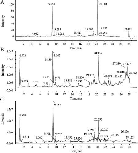
Table 1. Identification details of the components in SYE using UFLC-Q-TOF-MS/MS technique.
Figure 2. Chemical structural formulas of the identified constituents in SYE. Six constituents were confirmed by retention time and MS data of reference substances, and 23 constituents were characterized by MS data and related studies.
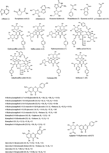
Figure 3. Time for arteriole occlusion of thrombus induced by laser irradiation in mice vessels. SYE was applied at three doses of 13, 26 and 52 mg/kg body weight by intraperitoneal injection on C57BL/6J mice. After 30 min, thrombus formation was induced in brain arterioles by laser irradiation and the time necessary for arteriole occlusion was measured. SYE was compared with heparin sodium injection (0.2 mL/kg). The graph depicts the mean values ± SD (n = 6); *p < 0.05 and **p < 0.01 as compared with the control.
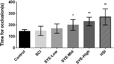
Figure 4. Arteriole diameter measurement during laser irradiation in the control (A) and SYE (B) group with high dosage. A brain arteriole of C57BL/6J mice with diameter of 18 ± 3 μm was selected as the target vessel for occlusion and was zoomed in at 20× magnification under TPLSM.
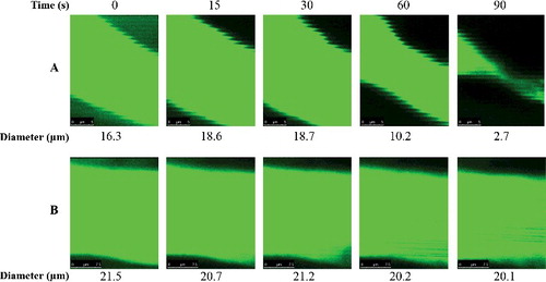
Figure 5. Ratio of vessel diameter. SYE was applied at three doses of 13, 26 and 52 mg/kg body weight by intraperitoneal injection on C57BL/6J mice. After 30 min, thrombus formation in brain arteriole was induced by laser irradiation and the diameter changes were measured and calculated as the ratio of diameter change to the initial diameter. The graph depicts the mean values ±SD (n = 6); *p < 0.05 and **p < 0.01 as compared with the control.
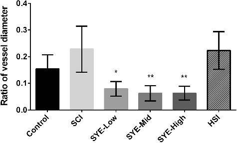
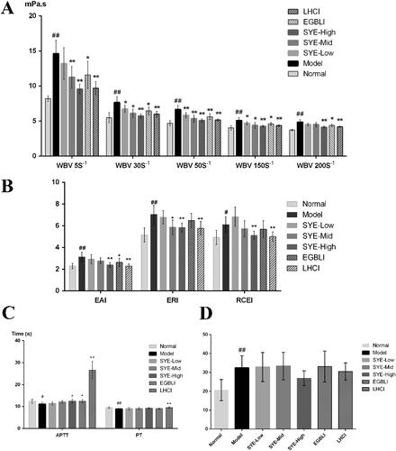
Figure 6. Hemorheology parameters measurements: WBV (A) at different shear rates; EAI, ERI and RCEI (B); APTT and PT (C); MPAR (D). SYE was applied at three doses of 1.5, 3.0 and 4.5 mg/kg body weight by intramuscular injection on SD rats. The graph depicts mean values ± SD (n = 8); #p < 0.05 and ##p < 0.01 as compared with the normal, *p < 0.05 and **p < 0.01 as compared with the model.