
Figures & data
Figure 1. Fractionation and purification of haemocyanin. (A) Fractionation of E. verrucosa dissociated haemocyanin chromatographed on a Fast Flow Sepharose Q column. Insert: Electrophoretic patterns of isolated fractions on a 10% SDS PAA gel electrophoresis (peaks 2, 3, 4 and 5). (B) HPLC purification of structural subunit 5 (SU5) in a Nucleosil 100 RP C18 column. (C) MALDI spectrum of structural subunit 5 (SU5) from E. verrucosa haemocyanin isolated on an anion exchange Fast Flow Sepharose Q column. The sample was measured by MALDI-TOF Ultraflex II.
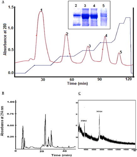
Figure 2. Carbohydrate analysis of haemocyanin subunits. (A) Orcinol–sulphuric acid test of E. verrucosa haemocyanin subunits eluted by HPLC and applied on a silica-gel plate. The spots on lane I are: 1 (2 mg/mL mannose); 2 (1 mg/mL mannose); 3 (0.5 mg/mL mannose); 4 (0.25 mg/mL mannose); 5 (0.1 mg/mL mannose). The spots on lane II are: 1 – SU1; 2 – SU2; 3 – SU3; 4 – SU4; 5 – SU5. (B) Carbohydrate and protein content of haemocyanin structural units of E. verrucosa haemolymph.
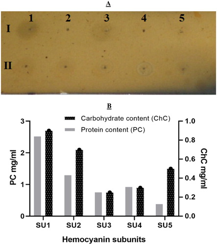
Figure 3. Antibacterial activity of native haemocyanin and its fractions. (A) Antibacterial activity of native haemocyanin (EvH) and its five fractions (numbers 1–5 represent subunits SU1–SU5, respectively) against different pathogenic bacteria: A/Bacillus subtilis; B/Escherichia coli; C/Salmonella enterica; D/Staphylococcus epidermidis. (B) MIC of subunit 1 (SU1) on test microorganism Staphylococcus epidermidis: 1 – 50 µg/mL; 2 – 25 µg/mL; 3 – 12.5 µg/mL; 4 – 6.25 µg/mL; 5 – 3.125 µg/mL; 6 – 1.562 µg/mL.
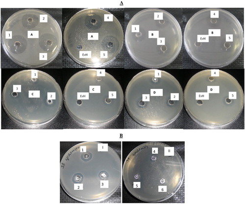
Table 1. Antibacterial activity of E. verrucosa haemocyanin and its subunits based on zones of inhibition (mm).
Table 2. Minimum inhibitory concentration MIC (µg/mL) and minimum bactericidal concentration MBC (μg/mL) of haemocyanin structural units on test microorganisms.