
Figures & data
Figure 1. M. communis plant (A), dried leaves (B) and extract (C).
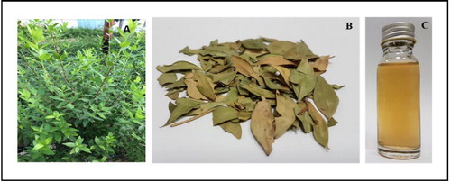
Figure 2. Flasks before and after reacting with silver nitrate (A), UV-Visible spectrum of the silver nanoparticle and capping of nanoparticle surface (B) and Zeta potential of nanoparticles (C).
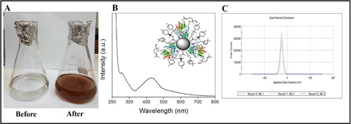
Figure 3. TEM micrograph (A–C) and EDAX of the silver nanoparticles (D).
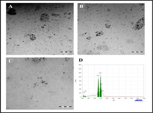
Figure 4. XRD analysis (A) and FTIR measurements of silver nanoparticles (B).
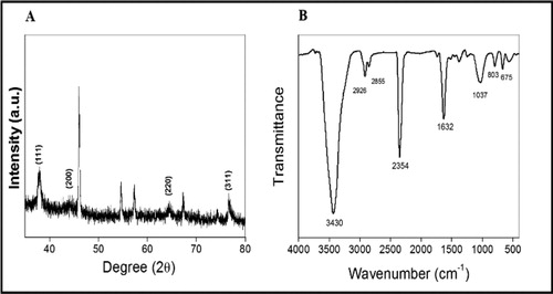
Figure 5. Antibacterial activity of silver nanoparticles against methicillin-resistant S. aureus (A) and E. coli (B).
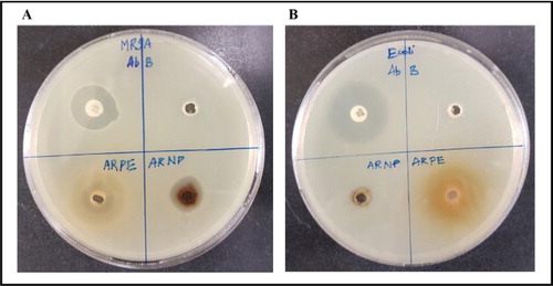
Table 1. Zone of inhibition of plant extract (ARPE) and nanoparticle (ARNP) against methicillin-resistant S. aureus (MRSA) and E. coli.
Table 2. MIC and MBC of silver nanoparticle against E. coli and methicillin-resistant S. aureus (MRSA).
Data availability
The data used to support the findings of this study are included in the article. Any specific details will be delivered by corresponding author upon request.