Figures & data
Figure 1. MDA (A) and tGSH (B) levels in the liver tissues of SHAM, LIR and BLIR groups.
Note: n = 6; *p < 0.001 vs. SHAM group; **p < 0.001 vs. LIR group; α, p > 0.05 vs. SHAM group.
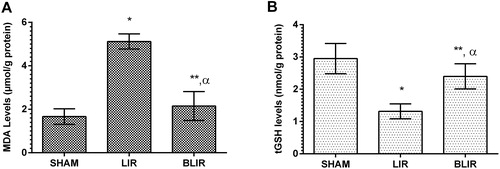
Figure 2. COX-1 (A) and COX-2 activities in the liver tissues of SHAM, LIR and BLIR groups.
Note: n = 6; *p < 0.001 vs. SHAM group; **p < 0.006 vs. LIR group.
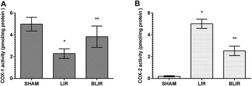
Figure 3. ALT (A) and AST (B) activities in the blood samples of SHAM, LIR and BLIR groups.
Note: n = 6; *p < 0.001 vs. SHAM group; **p < 0.001 vs. LIR group; α. p > 0.05 vs. SHAM group.
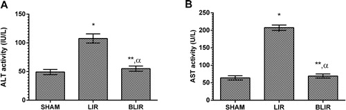
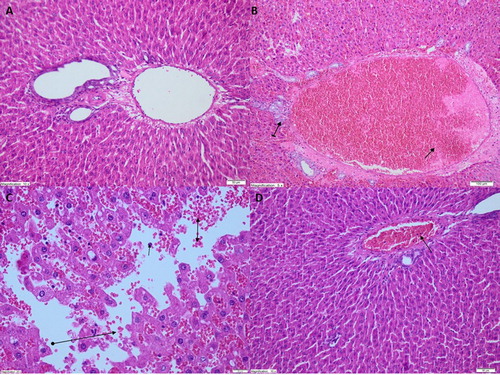
Figure 4. Light microscope images of liver tissue in SHAM (A) group, LIR group (B,C) and BLIR group (D).
Note: A. Normal view of liver tissue structure in SHAM animal group (HEX200), B. Dilated congested blood vessels (straight arrow) and increased bile duct proliferation (tailed arrow) were observed in liver tissue of LIR group (HEX100), C. Polymorphonucleated leukocytes (straight arrows), edema and hemorrhage (bilateral arrows), and destruction (round tailed arrows) were observed in liver tissue of LIR group (HEX400), D. Near-normal appearance of liver tissue in BLIR group except for progressing dilated blood vessels (HEX200).