
Figures & data
Figure 1. Agarose gel electrophoresis (1% agarose) of PCR amplified products (size 650 bp) using ITS1 and ITS4 PCR primer sets. Lane 1 is C. cladosporioides isolate BOU1 and Lane M is DNA marker (1 kb, Thermo Scientific).
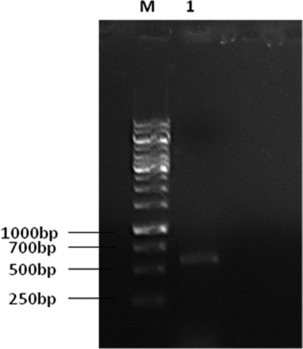
Figure 2. Isolated C. cladosporioides, strain BOU1: (A) infected brown planthopper (BPH) heavily colonized with BOU1; (B) growth of the infecting fungus from BPH in PDA medium at 27 °C for 7 days; (C) isolated fungal spore from the plate shown in B; (D) mycelial growth of fungus from a single spore in PDA after 24 h of incubation at 27 °C; and (E) mycelial growth of the isolated fungus at day two at 27 °C in water agar medium.
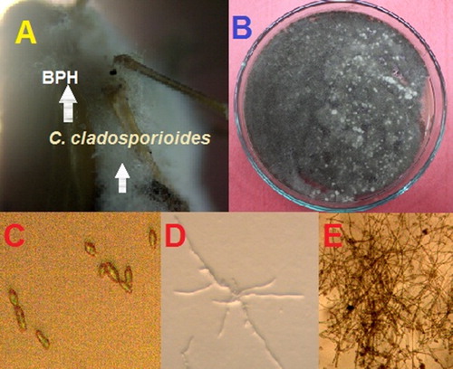
Figure 3. A phylogenetic tree constructed based on neighbor-joining method using MEGA 4.0 software. ITS1 and ITS2 regions of the seven isolates- BOU1 (This article), BFM-L47, EKF1, ZJDF16, OKF2, N37ZS and MC-24-L of Cladosporium spp. were aligned.
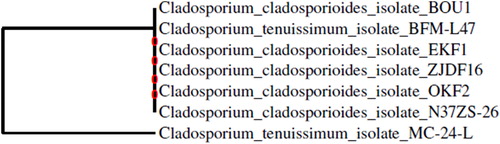
Figure 4. Colony area of C. cladosporioides, strain BOU1 in various media: (A) potato dextrose agar (PDA); (B) potato dextrose agar with yeast (PDAY); (C) sabouraud dextrose agar (SDA) and (D) synthetic nutrient poor agar (SNA) media.
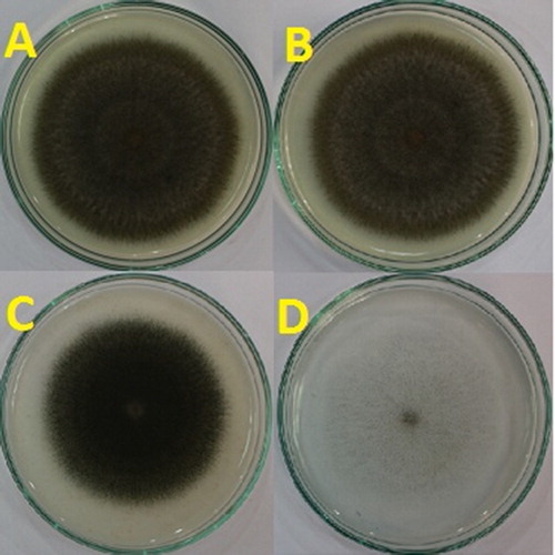
Table 1. Growth and enzyme activities of C. cladosporioides, strain BOU1 in different culture media.
Figure 5. Mortality rate (mean ± standard error [SE]) of whitefly after application of C. cladosporioides, strain BOU1.
Note: n, number of whitefly nymphs; same letters indicate statistically non-significant differences (LSD-test, following one-way ANOVA: P < 0.05).
![Figure 5. Mortality rate (mean ± standard error [SE]) of whitefly after application of C. cladosporioides, strain BOU1.Note: n, number of whitefly nymphs; same letters indicate statistically non-significant differences (LSD-test, following one-way ANOVA: P < 0.05).](/cms/asset/85fce333-5e3a-495e-bdaa-92f7968dd130/tbeq_a_1695541_f0005_c.jpg)