
Figures & data
Figure 1. dCATH mRNA level in different tissues at post-hatch day 1 (a), day 7 (b), day 14 (c), day 21 (d) and day 28 (e).
Note: β-actin was used to normalize the target gene expression profiles. Data relate to 3 healthy ducks/group and are presented as means ± SD

Figure 2. Purity and molecular weight of the dCATH peptide analyzed by HPLC (a) and ESI-MS (b), respectively.
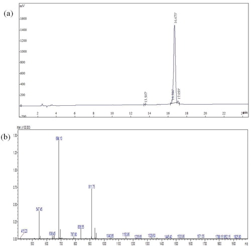
Figure 3. Antiserum titer of immuned mouse against the dCATH peptide.
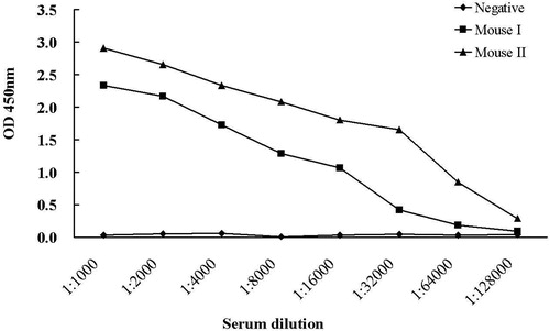
Table 1. Cell fusion and screening of hybridoma clones against dCATH.
Figure 4. Analysis of the purified dCATH mAb. (a) Titer of the mAb determined by iELISA. The minimum OD value greater than 1 is at a titer of 1.28 × 10⁵, indicating that the antibody remained active after purification. Negative control was ascites from negative mouse. Three replicates were performed for each test. (b) The isotype of dCATH mAb determined by isotyping kit (IgA, IgG1, IgG2a, IgG2b, IgM, Lambda, Kappa). (c) SDS-PAGE analysis of the dCATH mAb. Lane M: Marker (RP1400, Solarbio Life Science®, Shanghai, China), lane 1: the purified mAb.
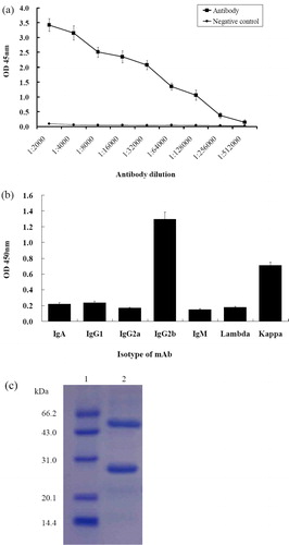
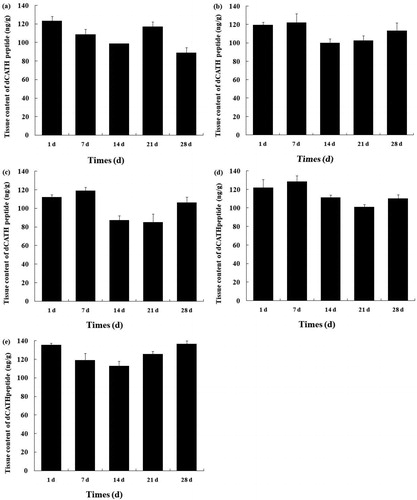
Figure 5. Tissue concentration of the dCATH peptide in healthy ducks. Liver (a), kidney (b), spleen (c), pancreas (d) and bursa (e) sampled from healthy ducks at day 1, day 7, day 14, day 21 and day 28 after hatching. Data relate to 3 healthy ducks/group and are presented as means ± SD.
Data Availability Statement
The data that support the findings of this study are available from the corresponding author, Xingjun Feng, upon reasonable request.