Figures & data
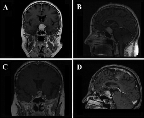
Figure 1. Case 1. Preoperative coronal (A) and sagittal (B) contrast-enhanced T1-weighted MR images showing a large TSM with superior extension. Postoperative coronal (C) and sagittal (D) contrast-enhanced T1-weighted MR images showing GTR of the TSM.
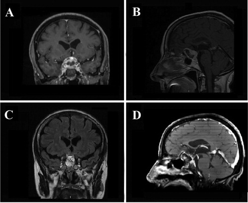
Figure 2. Case 2. Preoperative coronal (A) and sagittal (B) contrast-enhanced T1-weighted MR images showing a large TSM with inferior sellar extension and midline location. Postoperative coronal (C) and sagittal (D) contrast-enhanced T1-weighted MR images showing GTR of the TSM.
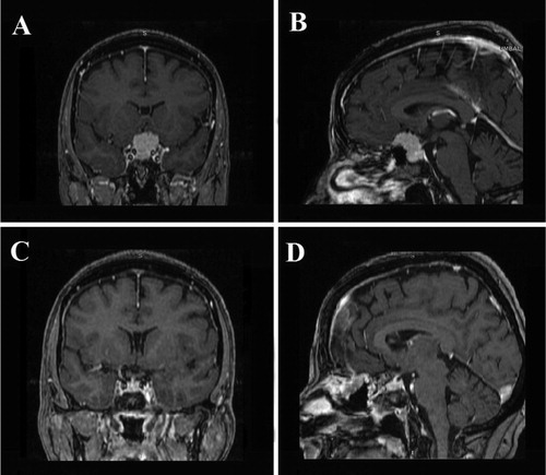
Figure 3. Case 3. Preoperative coronal (A) and sagittal (B) T2-weighted MR images showing a large TS/PS tumor without lateral extension beyond the internal carotid arteries. Postoperative coronal (C) and sagittal (D) contrast-enhanced T1-weighted MR images showing GTR of the TSM.
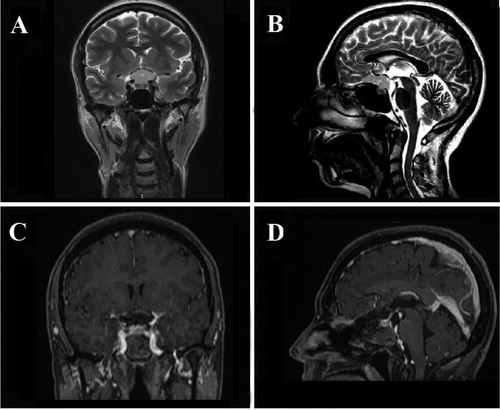
Figure 4. Case 4. Preoperative coronal (A) and sagittal (B) contrast-enhanced T1-weighted MR images showing a large midline TSM adherent to the brain structures and with mainly suprasellar extension. Postoperative coronal (C) and sagittal (D) contrast-enhanced T1-weighted MR images showing STR of the TSM.