Figures & data
Table 1. Specimen numbers.
Figure 1. Box plot of six basilar artery specimens of mice in GSE46696.
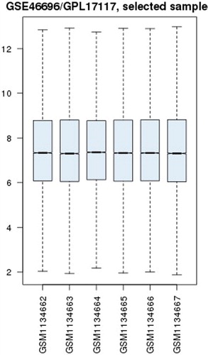
Figure 2. Unsupervised hierarchical clustering analysis of six basilar artery specimens in GSE46696.
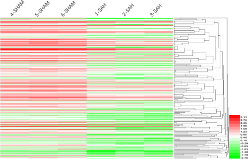
Figure 3. Functional enrichment analysis of differentially expressed genes in basilar artery specimens of mice in GSE46696.
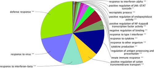
Figure 4. KEGG pathway of differentially expressed genes.
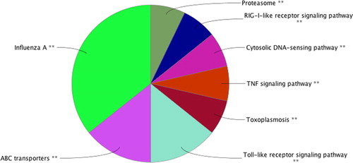
Figure 5. Interaction network diagram of genes associated with cerebral vasospasm after subarachnoid haemorrhage.
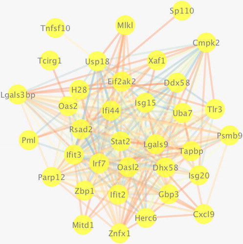
Figure 6. Network diagram of genes involved in immune response regulation.
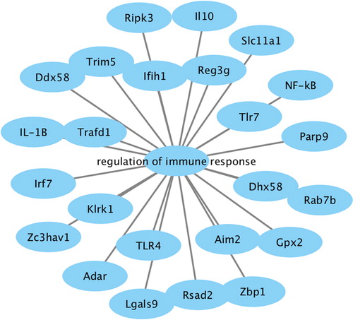
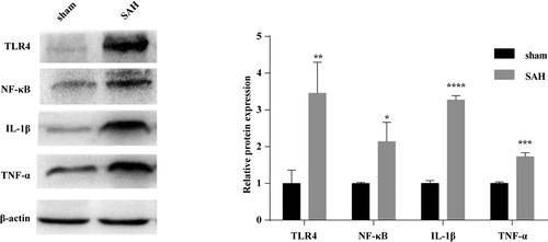
Figure 7. Western blotting analysis of IL-1β, TNF-α, NF-κB and TLR4 in brain tissues of SAH mice. Three days after model establishment, the expression levels of inflammatory cytokines IL-1β, TNF-α and TLR4/NF-κB inflammatory signalling pathway-related proteins, NF-κB, TLR4 were statistically higher in the brain tissues of mice in the SAH model group than in the sham group (all p < 0.05).