
Figures & data
Figure 1. CTPA (A) and P-SPECT/CT (B) of Case 1. (A) CTPA of pulmonary artery, main, lobar and segmental branches without visible defects of filling. In the area of left lung lingula parenchyma consolidation and filling defect in the level of subsegmental branches. Small pleural effusion on the left. (B) Perfusion SPECT/CT in the left lung showing perfusion defect in lung lingula with parenchyma consolidation. Small pleural effusion. In the right lung hypoperfusion defects in small branches in lower lobe.
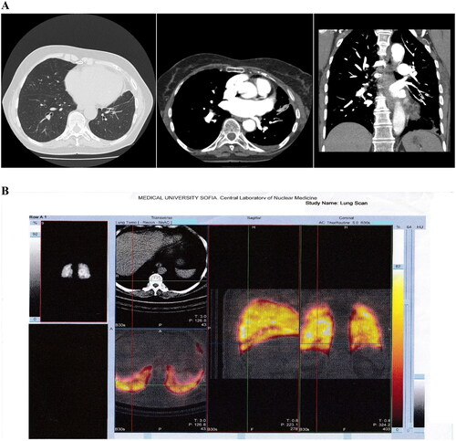
Figure 2. CTPA (A) and P-SPECT (B) of Case 2. (A) CTPA with no filling defects in pulmonary artery, its main, lobar and segmental branches, which are of normal size. (B) Perfusion SPECT/CT showing both side hypoperfusion in the subsegmental level of pulmonary artery.
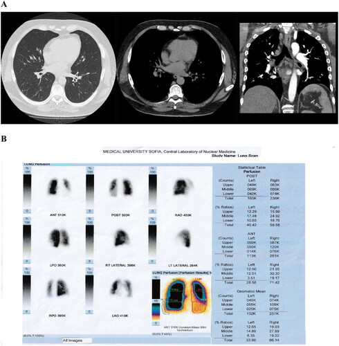
Table 1. Crosstable of imaging methods.
Table 2. Diagnostic value of the imaging methods.
Data availability statement
The data that support the findings of this study are available from the corresponding author [Sevda Naydenska], upon reasonable request.