
Figures & data
Table 1. Blood tests during hospitalization.
Table 2. CSF analysis on admission (on day 1) and control LPs (on days 7 and 17).
Table 3. Cytokines levels in CSF and serum specimen.
Figure 1. Control CT of the brain. No pathological changes in the parenchyma. Presence of anatomic variation (smaller left sinus transverses).

Figure 2. Chest radiography. Inflammatory pneumonitis in the right basal lung. Hypoventilated surrounding parenchyma. Cardiac shadow with elongated left ventricular arch.
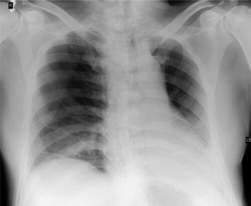
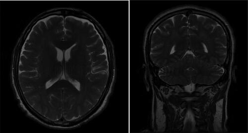
Figure 3. Brain MRI transverse and coronal plane.
Data availability statement
The anonymized raw clinical and laboratory data associated with the present report are available from the corresponding author [P.A.] upon a reasonable request.