Figures & data
Figure 1. Two types of lateral root, e.g. root of Puluik Arang under 10% PEG treatment.
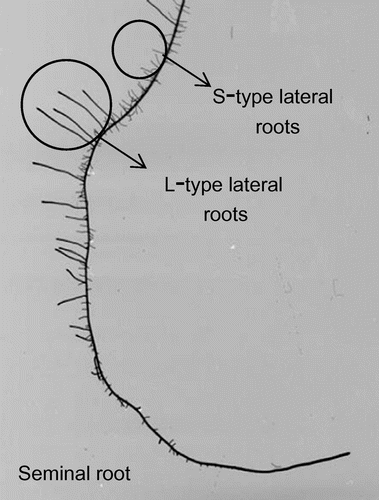
Table 1. Water uptake, dry matter production and root morphological traits of two rice cultivars in response to PEG stress.
Figure 2. Comparison of the copy numbers of expressed aquaporin genes in the seminal roots and lateral roots emerged from seminal root of different cultivars under osmotic stress. Roots of 13-day-old plants were sampled three hours after lights-on. Three replicates of each cultivar for each treatment were conducted using different plant samples. Bars indicate the standard error (n = 3). Asterisk represents a significant difference between cultivars (p < 0.05, t-test). Probability from the results of ANOVA was shown to determine the individual and interaction effects of the treatment and cultivar.
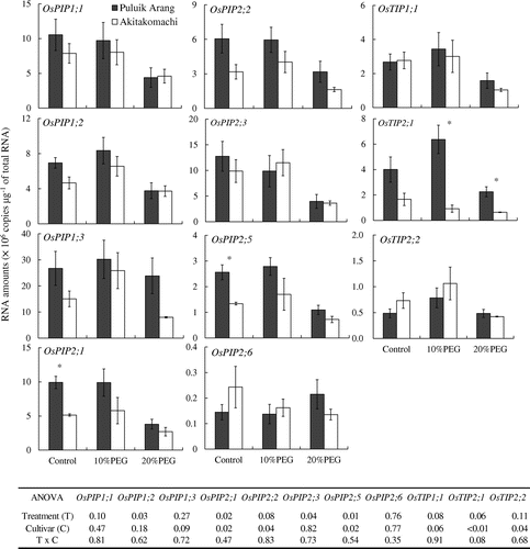
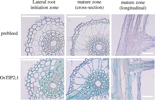
Figure 3. Immunolocalization of OsTIP2;1 in seminal root tissue of the cultivar ‘Puluik Arang’ subjected to 10% PEG treatment. Root tissue samples from 13-day-old plants were excised from the lateral root initiation zone (approximately 1–2 cm from the root tip) and from the mature zone (middle position of seminal root) of the seminal root. Bars represent 100 μm.