Figures & data
Figure 1. OS in rat skin (a) markers of OS: free radicals accumulation (MTT), XO activity (AXO), and lipid peroxidation (MDA); control group (Control), SSUV-exposed group (SSUV); propylthiouracil-induced hypothyroid group (PTU), and propylthiouracil-induced hypothyroid group, exposed to SSUV radiation (PTU + SSUV). (b) Formation of MDA as a function of radical accumulation in the skin of: control group (С); irradiated group (UV); propylthiouracil-induced hypothyroid group (PTU), and irradiated hypothyroid group (PTU&UV).
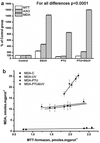
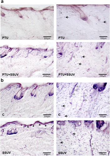
Figure 2. NADPH-d reactivity in hypothyroid (PTU) (a), and euthyroid (C) (b) rat’s skin, with or without SSUV exposure. Black arrows indicate dermal fibroblasts, black arrowheads – keratinocytes in epidermis, white arrowheads – epithelial cells of hair root follicles, white arrows – blood vessels. The positive cells are visualized with dark blue color.
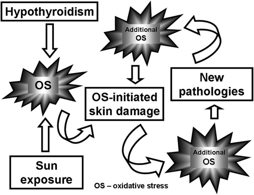
Scheme 1. Effect of the sunlight exposure on the OS and OS-induced pathologies in hypothyroid rats.
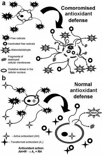
Scheme 2. Effect of the free radicals on attack on the lipid peroxidation of the cell: (a) if the antioxidant defense is not properly working, (b) if the antioxidant defense is normal.