Figures & data
Table 1. Forward and reverse primers sequence for candidate genes.
Table 2. Body weight, Lee's index, fasting serum glucose, insulin levels and HOMA-IR.
Table 3. Serum lipid profile among studied groups.
Table 4. Serum reproductive hormones among studied groups.
Figure 1. Serum tumour necrosis alpha (TNF-α) level: Data were expressed as mean ± SD, aSignificant change vs. control, bSignificant change vs. PCOS-IR, cSignificant change vs. MET, dSignificant change vs. SeNPs, eSignificant change vs. SeNPs + MET. PCOS, Polycystic ovarian syndrome; MET, Metformin; SeNPs, Selenium nanoparticles.
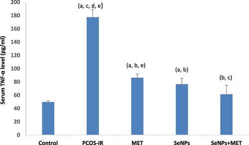
Figure 2. Oxidative stress markers in ovarian tissues: (A) The MDA level (nmol/mg protein). (B) SOD activity (U/mg protein). (C) GSH level (μmol/g protein). Data were expressed as mean ± SD, aSignificant change vs. control, bSignificant change vs. PCOS-IR, cSignificant change vs. MET, dSignificant change vs. SeNPs, eSignificant change vs. SeNPs + MET. PCOS, Polycystic ovarian syndrome; MET, Metformin; SeNPs, Selenium nanoparticles.
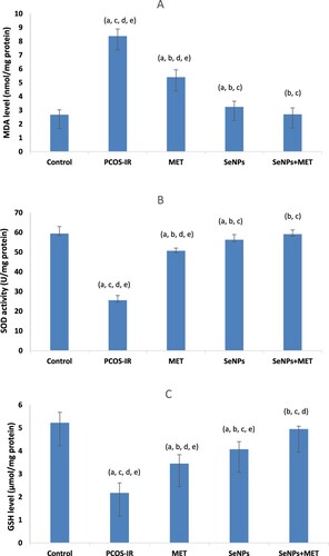
Table 5. Mitochondrial function parameters among studied groups.
Figure 3. Quantitative Real-time PCR analysis of mRNA expression levels of PI3K gene in ovarian tissue among the studied groups. Data were expressed as mean ± SD, a significant change vs. control, b Significant change vs. PCOS-IR, c Significant change vs. MET, d Significant change vs. SeNPs, e Significant change vs. SeNPs + MET. PCOS, Polycystic ovarian syndrome; MET, Metformin; SeNPs, Selenium nanoparticles.
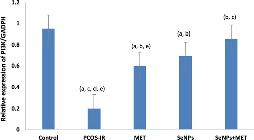
Figure 4. Quantitative Real-time PCR analysis of mRNA expression levels of Akt gene in ovarian tissue among the studied groups. Data were expressed as mean ± SD, a significant change vs. control, b Significant change vs. PCOS-IR, c Significant change vs. MET, d Significant change vs. SeNPs, e Significant change vs. SeNPs + MET. PCOS, Polycystic ovarian syndrome; MET, Metformin; SeNPs, Selenium nanoparticles.
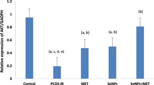
Figure 5. Histopathological examination of the ovary in different groups: A) Control group showing the antral follicle (thick arrow) and corpus luteum (CL) (Figure A1). The follicle shows healthy granulosa cells, zona pellucida (double arrow) and oocyte (thin arrow) within the follicular antrum (FA) (Figure A2). B) PCOS-IR model group showing multiple follicular cysts (stars) with absent corpus luteum. C) MET-treated group showing the antral follicle (thick arrow) with oocyte (thin arrow) and its follicular antrum. The corpus luteum (CL) reappears but with irregular luteal cells compared to the control. A cystic follicle (star) is also seen. D) SeNPs-treated group showing antral follicle (thick arrow) with oocyte (thin arrow) but with scanty follicular antrum (FA). The corpus luteum (CL) reappears but with irregular luteal cells. E) SeNPs + MET-treated group showing multiple primary follicles (white arrows) (Figure A1). The antral follicle (thick arrow) with oocyte (thin arrow), zona pellucida (double arrows) and its follicular antrum (FA). The corpus luteum (CL) is also seen (Figure A2). All slides are H&E stained. Magnification: × 200.
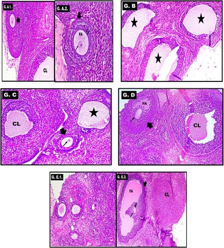
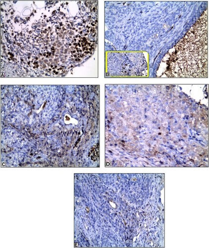
Figure 6. Immunohistochemical expression of ki-67 in different groups: A) Control group showing strong immunopositive staining in granulosa cells. B) PCOS-IR model group showing Ki-67 nuclear expression mainly in the theca cells and nearly absent in granulosa cells (higher power figure is inserted in the left box). C) MET-treated group showing ki-67 expression mainly in granulosa cells. D) SeNPs -treated group showing ki-67 expression mainly in granulosa cells. E) SeNPs + MET-treated group showing higher ki-67 expression and is present mainly in granulosa cells. Magnification: × 400.