Figures & data
Table 1. Serial neuropsychological examinations
Figure 1. Serial MRI results. *Abbreviation: MRI: Magnetic resonance imaging (MRI). Overview of raw data of MRI scans of the brain which were performed between 2000 and 2009, including scanner type and age at time of performance. At visual inspection there is no progressive atrophy
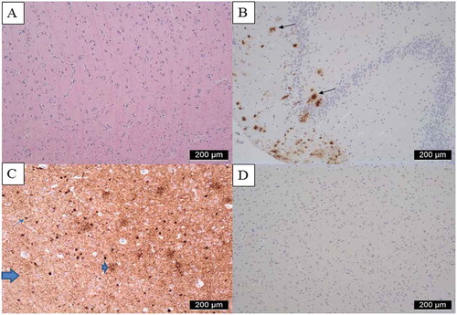
Figure 2. Neuropathological images of brain autopsy. *Abbreviations: ABC score; Amyloid βeta plaque score (A), Braak neurofibrillary tangles (NFT) (B), Consortium to Establish a Registry for AD (CERAD) neuritic plaque score (C). Neuropathological examination revealed a A3 B2 C2 score fitting an intermediate score for Alzheimer’s disease neuropathological change (Thal et al., Citation2002). A. Hematoxylin and eosin staining (HE) of second frontal gyrus displaying normal cellularity B Amyloid beta staining of hippocampus, Cornu Ammonis (CA) area 4, showing amyloid plaques Thal phase 4 out of 5 (A3)C AT8 staining of transentorhinal cortex, showing moderate presence of phosphorylated tau (NFT stage 2 (B2) as tangles (example thin arrow); neuropil threads (thick arrow) and as neuritic plaques (example arrow head). D Negative TDP-43 staining of second frontal gyrus