
Figures & data
Figure 1. Steps of the Mefix procedure. (A) Schematic diagram of Petersen’s space (purple dotted line), jejunal mesentery (blue area, jejunal side, at site 30 cm distal J-J) with transverse mesocolon (red area, colonic side). (B) Exposure of the suture area jejunal side & colonic side (red line, SMA vascular arcade) (Step 1). (C) Step 2. The suture started between jejunal side just below of SMA vascular arcade and colonic side. (D,E) Step 3. To anchor the mesentery, two-point backward sutures were performed, and Mefix was done.
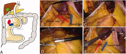
Table 1. Patient demographics.
Table 2. Clinicopathologic comparison between Petersen closure group (closure) and distal mesentery fixing group (Mefix).
Supplemental material