Figures & data
Figure 1. Precontrast (a), postcontrast (b), and subtraction (c). T1-weighted MR images of the left eye showing contrast enhancement of the tumor (white arrows in b) and distal optic nerve (black arrow in b). Enhancement of the optic nerve (black arrow in b and c) reached 3 mm from the lamina cribrosa into the optic nerve and reached the outer edges (width of the postlaminar enhancement was about 3.5 mm). Temporal from the tumor the retina has detached and shows T1-hyperintense subretinal fluid (white arrowhead in b), but no enhancement (no signal on the subtraction image, c).
Figure 2. Histology slides showing an enlarged distal optic nerve with tumor cells (purple) occupying the entire width of the optic nerve (black arrows in a) corresponding with findings on MRI. Notice the lamina cribrosa bulging outwards (white arrow in a), probably from a high-ocular pressure (44 mmHg), and possibly partly due to mass effect from the intraocular tumor (§ in a). The tumor cells (black arrows in b) reach the outer layers of the optic nerve all the way up to the pia mater (* in b) with the CSF space represented by the void adjacent to the pia († in b).
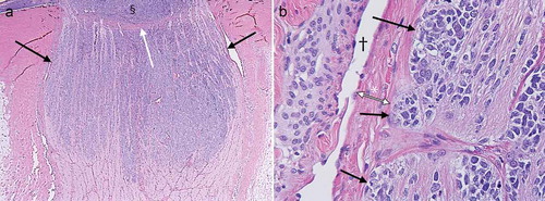
Figure 3. Maximum diameters on axial and oblique coronal reconstructions of FIESTA images (pixel size is 0.27 mm and slice thickness 0.6 mm) of the distal optic nerve of the unaffected OD (a and c) and affected OS (b and d).