
Figures & data
Table 1. Demographic data on children with microphthalmia and anophthalmia including age at examination, completed gestational age and birth weight, mode of delivery, diagnosis, unilaterality or bilaterality and microphthalmia subgroup
Figure 1. (a–d) Girl born with total blindness (amaurosis) due to right sided anophthalmia and left sided microphthalmia. There was no bulb on the right side and a very small, rudimentary bulb 5 mm on the left side. (a) New-born child, one month of age. (b) Treatment with expanding conformers, here 10 months of age. (c) Six years of age with bilateral prostheses. (d) MRI of the brain and orbits showing anophthalmia right eye and microphthalmia left eye (Published with permission from parents) .
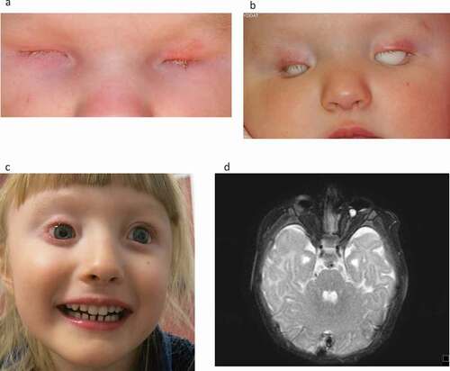
Figure 2. (a–c) Child born with small microphthalmic left eye. (a) Two months of age. (b) Treated with expanding conformers-at eight months of age she is wearing a-left eye prosthesis. Right eye normal. (c) Magnetic resonance tomography (T-2 weighted) of the brain and orbits. Note the microphthalmic left eye. (Published with permission from the parents).
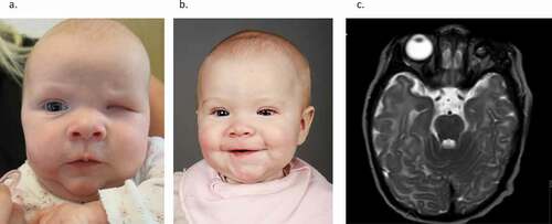
Figure 3. Prostheses from one child with bilateral anophthalmia.
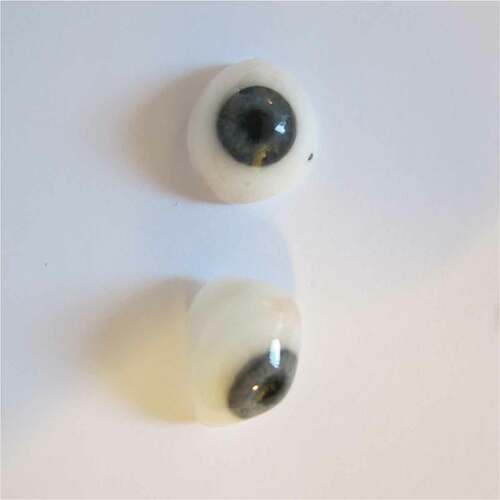
Table 2. Somatic comorbidities in children with anophthalmia and microphthalmia including subgroup, isolated or non-isolated A/M, genetic findings, magnetic resonance imaging of the bulb and CNS, and disorders in different organ systems
Table 3. Ocular characteristics including diagnosis, visual functions, VEP, intraocular pressure, treatment with expander (E) and/or prosthesis (P)
Figure 4. Somatic co-morbidities in patients with anophthalmia or microphthalmia.
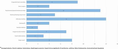
Table 4. Health-related quality of life reported by parents and children according to age