
Figures & data
Table 1. Genetic and clinical data
Table 2. Disease-associated alleles reported to date
Figure 1. Location and features of disease-associated RCBTB1 variants.
The schematic figure shows the location of all the disease-associated variants in RCBTB1 described in the literature and in this report. Novel variants are shown in bold font.
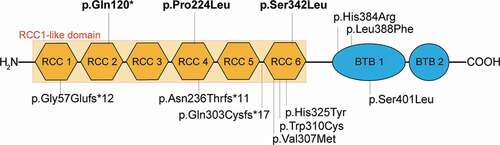
Figure 2. Fundus imaging of patients with RCBTB1-associated retinal dystrophy.
Fundus photography (A-C), fundus autofluorescence (FAF; D-F), and SD-OCT (G-I) were obtained in all patients. In Patient 1, nummular patches of fovea-abutting atrophy were observed on exam and with fundus photography (A). Subtle pigment mottling was present in the midperipheral retina. FAF (D) highlighted the nummular nature of the atrophy and also showed generalized macular hypofluorescence without prominent peripheral disruption. SD-OCT (G) delineated areas of atrophy with a restricted Island of foveal sparing in addition to showing more widespread RPE irregularities.In Patient 2, fundus exam and photography (B) showed fovea-sparing macular atrophy. Fine pigment deposits, nummular areas of chorioretinal atrophy, and occasional larger pigment clumps were present in the peripheral retina. FAF (E) further illustrated the distribution of atrophy and pigment disruption. SD-OCT (H) showed generally intact foveal lamination. Areas of atrophy were also present with associated cystoid spaces and outer retinal tubulations (ORTs).In Patient 3, fundus exam and photography (G) showed extensive macular atrophy with several prominent pigment clumps. Fine pigmentary deposits in the peripheral retina formed a reticular pattern in are``as. FAF (H) showed coalescing nummular hypofluorescence in the macula with narrow strips of preserved signal. Reduced hypofluorescence was present throughout the midperiphery. Extensive chorioretinal atrophy and ORTs were present on SD-OCT (I) with limited areas of preserved outer retinal structure.
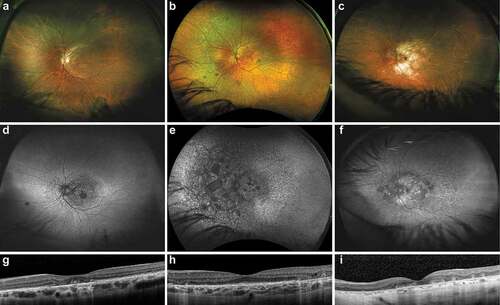
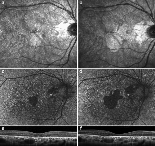
Figure 3. Evolution of macular findings in a single patient with RCBTB1-associated retinal dystrophy.
Progressive macular disease was seen over a 3 year interval in Patient 1 as evaluated in near-infrared (A,B), fundus autofluorescence (C,D), and SD-OCT (E,F) imaging, presented for the right eye.
Supplemental material