Figures & data
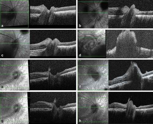
Figure 1. SD-OCT imaging of a retinal retinoblastoma relapse following epiretinal seed implantation. (a) Scan over the inferior parapapillary region at the end of the first-line treatment showing an intact retina. (b) Same region at time of the vitreous relapse showing a large epiretinal seed anchored and locally disrupting the inner limiting layer. (c) Complete regression of the epiretinal seed after four combined intravitreal melphalan and topotecan. (d) Isolated intra-retinal relapse 8 months later originating from the site of the inner limiting layer invasion at time of the vitreous seeding. (e) Flat retinal scar post thermotherapy.
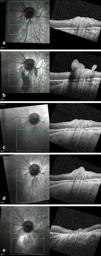
Figure 2. SD-OCT imaging of an optic nerve retinoblastoma relapse following an epipapillary seed implantation. (a) Scan of the optic disc at the end of the first-line treatment showing an intact optic nerve head. (b) Same imaging at time of the vitreous relapse showing an epipapillary seed filling the optic nerve cup. (c) Regression of the epipapillary seed after two intravitreal melphalan. (d) Massive relapse masking the optic nerve 2 months later, with no demarcation line between the anchored seed and the papilla. (e) Regression of the relapse after 2 intraarterial and 4 intravitreal injections with combined topotecan and melphalan. (d) New epipapillary growth with suspicion of prelaminar invasion four months later. (g) Regression of the relapse after two cycles of combined intra-arterial melphalan and topotecan. (h) Infraclinical optic nerve head recurrence 6 months later motivating secondary enucleation.