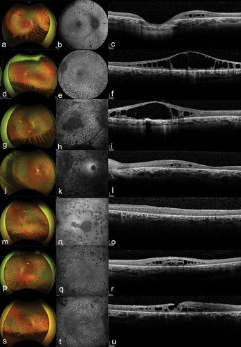
Figure 1. Typical findings in patients with mild Zellweger spectrum disorder (ZSD) with biallelic variants c.2528 G>A (p.Gly843Asp) in PEX1 on fundus photographs, fundus autofluorescence (FAF), and spectral-domain optical coherence tomography (SD-OCT).
All patients had a hyperautofluorescent speckle-pattern on FAF (b, e, h, k, n, q, t) and varying degrees of integrity loss of the external limiting membrane (ELM) and ellipsoid zone (EZ) on SD-OCT (c, f, i, l, o, r, u).
A, B, C, Right eye of a 25-year-old patient (A1) with on fundus photos (a) macular atrophy, bone-spicule hyperpigmentation and mild attenuation of retinal vasculature and on SD-OCT (c) integrity loss of the ELM and cystoid fluid collections in the outer nuclear layer (ONL).
D, E, F, Left eye of a 20-year-old patient (A2) with on fundus photos (d) round hyperpigmentations, and on SD-OCT (F) large cystoid fluid collections in the ONL with retinoschisis aspect.
G, H, I, Right eye of a 26-year-old patient (B1) with on fundus photos (g) bone-spicule and round hyperpigmentations and mild attenuation of retinal vasculature and on SD-OCT (i) large cystoid fluid collections in ONL with retinoschisis aspect.
J, K, L, Left eye of a 25-year-old patient (B2) with on fundus photos (j) bone-spicule and round hyperpigmentation with partial obliteration of retinal vasculature and on SD-OCT (l) loss of retinal lamination and parafoveal cystoid fluid collection in the ONL.
M, N, O, Left eye of a 17-year-old patient (C1) with on fundus photos (m) bone-spicule and round hyperpigmentations and on SD-OCT (o) loss of retinal lamination.
P, Q, R, Left eye of an 11-year-old patient (C2) with bone-spicule and round hyperpigmentations and mild attenuation of retinal vasculature on fundus photos (P) and on SD-OCT (R) central cystoid fluid collections in the ONL.
S, T, U, Right eye of a 44-year-old patient (E1) with on fundus photos (s) bone-spicule and round hyperpigmentations with partial obliteration of retinal vasculature and on SD-OCT (u) loss of retinal lamination and aspect of lamellar macular hole.
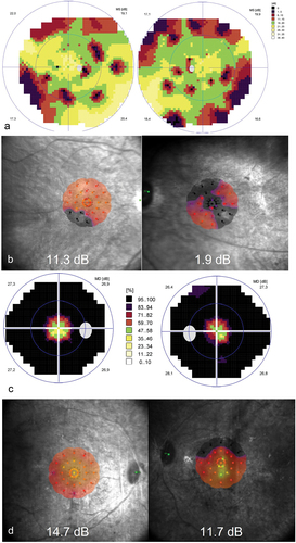
Figure 2. Typical findings on perimetry and microperimetry in patients with mild Zellweger spectrum disorder (ZSD).
(a) 90 degrees visual field measurement on Octopus 900 of patient A2 demonstrating peripheral constriction and mid-peripheral scotoma. Colour-coded heatmap reflects percentage deviation from healthy population.
(b) Heatmap of macular sensitivity of patient A2 on microperimetry, with grey loci reflecting areas of no sensitivity (absolute scotoma). Mean sensitivity is shown in white.
(c) 30 degrees visual field measurement on Octopus 900 of patient B2 demonstrating severe visual field constriction. Colour-coded heatmap reflects percentage deviation from healthy population.
(d) Heatmap of macular sensitivity of patient B2 on microperimetry, with grey loci reflecting areas of no sensitivity (absolute scotoma). Mean sensitivity is shown in white.
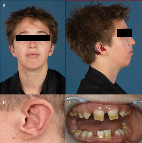
Figure 3. Mild craniofacial dysmorphic features and amelogenesis imperfecta in adults with mild Zellweger spectrum disorder (ZSD). Patients with comparatively mild ZSD had varying degrees of.
(a) low-set ears,
(b) attached earlobes, and
(c) amelogenesis imperfecta. Written informed consent was obtained from all patients for publication of these images.