Figures & data
Figure 1The chemical structures of the diterpenoids of Andrographis paniculata. Nees. Source: Matsuda et al. (Citation1994).
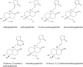
Table 1.. Cytotoxic activity (EC50 values, 72 h) of diterpenoid constituents of A. paniculata. and positive controls against a variety of cell lines.
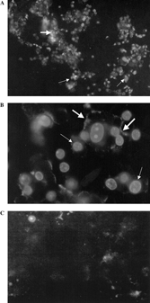
Figure 2The effect of (A) 14-Deoxy-11,12-didehydroandrographolide (EC50 values of 1.520 µg/ml, 72 h) (B) DNase I (1 U/ml), (C) etoposide (EC50 values of 1.9 µg/ml, 72 h), and (D) DMSO 1% (v/v) on T-47D cells as assayed with Deadend™ Colometric Apoptosis Detection System (Promega, USA). Arrows show darkly stained nuclei of T-47D cells indicating DNA fragmentation. No DNA fragmentation was observed in diterpenoid-treated cells. Trypan blue exclusion assay of the T-47D cells treated with 14-Deoxy-11,12-didehydroandrographolide (EC50 values of 1.5 µg/ml, 72 h) for (E) 24 h and (F) 72 h. Arrow shows necrotic cell.
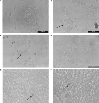
Figure 3The effect of 14-deoxy-11,12-didehydroandrographolide (EC50 values of 1.52 µg/ml, 72 h), on T-47D cell line as stained with Annexin-V-FLUOS™ kit (Roche, Germany). (A and B). The effect of 1% (v/v) DMSO, control vehicle on T-47D cell line. (C) Positive annexin V (thick arrows) and propidium iodide (thin arrows) stained cells were noted. Original magnification × 100 (A) × 400 (B, C).