Figures & data
Figure 1 Immunoblotting analysis of tyrosine protein phosphorylation in 0.1 mg/ml matrine-treated K562 cells at various times. Treatment of K562 with matrine resulted in an increase in tyrosine phosphorylation of proteins migrating at 220 (P220), 130 (P130), 90 (P90), and 69 (P69) kDa starting at 30 min after matrine addition and lasting for the entire experiment period. Phosphorylation of two proteins with 36 kDa and 34 kDa were downregulated, whereas both elevated to control levels by 24 h. Positions of molecular mass markers are shown to the right (in kilodaltons). Each experiment was repeated two times and similar results were obtained.
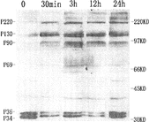
Figure 2 Immunoblotting analysis of the protein tyrosine phosphorylation of concentrations of matrine on K562 cells for 12 h. Exposure of K562 cells to concentrations of matrine induced an elevation in the tyrosine phosphorylation of proteins migrating at 220 (P220), 130 (P130), 90 (P90) and 69 (P69) kDa and a decrease of phosphorylation of two proteins with 36 kDa and 34 kDa. Elevated tyrosine phosphorylation of proteins declined somewhat the concentration of matrine increased, but changes in proteins with 36 kDa and 34 kDa remained stable. Positions of molecular mass markers are shown to the right (in kilodaltons). Each experiment was repeated two times and similar results were obtained.
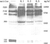
Figure 3 Immunoblotting analysis of the protein tyrosine phosphorylation of concentrations of STI571 on K562 cells for 12 h. Exposure of K562 cells to STI571 for 12 h resulted in a decrease in the tyrosine phosphorylation of the same proteins affected by matrine. Positions of molecular mass markers are shown to the right (in kilodaltons). Each experiment was repeated two times and similar results were obtained.
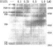
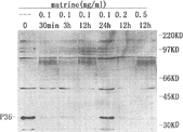
Figure 4 Immunoblotting analysis of the protein tyrosine phosphorylation in matrine-treated Raji cells. When 0.1 mg/ml of matrine was administrated to Raji cells, tyrosine phosphorylation of protein with 36 kDa was diminished dramatically at 30 min and almost disappeared at 12 h but elevated to the control level by 24 h. The striking declined phosphorylation was also found in Raji cells treated with 0.2 and 0.5 mg/ml matrine for 12 h. Positions of molecular mass markers are shown to the right (in kilodaltons). Each experiment was repeated two times and similar results were obtained.