Figures & data
Figure 1 Comparison of the growth inhibitory effects of two concentrations of tomatidine and solasodine alone and in combination (T + S) with that of cisplatin. HeLa, HT29, and MCF7 cells were exposed to 10 or 100 µM of cisplatin, tomatidine, solasodine, or the latter two combined for 48 h. Mean ± SD (n = 4). **p < 0.001; *p < 0.05 compared with control.
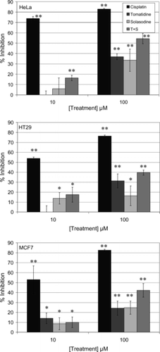
Table 1.. IC50 values for the test compounds as obtained from log dose response curves using GraphPad Prism.
Table 2.. Results of cell cycle analysis using nuclear PI staining.
Table 3.. Apoptosis results from annexin V–FITC/PI analysis.