Figures & data
Figure 2 Structure of baicalein (Mr = 270.23).
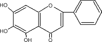
Figure 1 Samples screening for human apoA-I expression activators. Four different concentrations of samples were used for screening, each concentration tested in duplicate. (▵ represents 10 μ g/mL, ▴ represents 30 μ g/mL, ▪ represents 100 μ g/mL, and ▪ represents 300 μ g/mL. The abscissa represents five active samples obtained after the primary screening.)
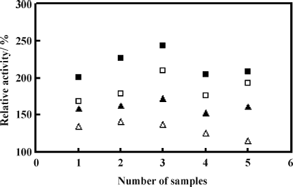
Figure 3 Dose-response of baicalein on human apoA-I promoter expression. Transactivation assay was performed as described in “Materials and Methods.” Data shown are mean ± SD tested in duplicate in the SApoA-I cell line. Relative activities are derived from: Baicalein reading/DMSO reading × 100%.
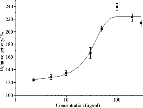
Figure 4 Effect of baicalein on ABCA1 promoter expression. Transactivation assay was performed as described under “Materials and Methods.” Data shown are mean ± SD tested in duplicate in the SABCA1 cell line. Relative activities are derived from: Baicalein reading/DMSO reading × 100%.
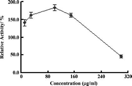
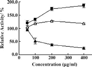
Figure 5 Baicalein increases apoA-I and apoA-II production and decreases apoC-III production in HepG2 cells. HepG2 cells were incubated with various concentrations of baicalein for 24 h, then the levels of apoA-I, apoA-II, and apoC-III protein were measured as described under “Materials and Methods.” Data are the means of triplicate wells, and the error bars indicate the range of triplicates. Relative activity is derived from: Baicalein reading/DMSO reading × 100%. ▪ represents apoA-I, ▵ represents apoA-II, and ▴ represents apoC-III.