1,688
Views
55
CrossRef citations to date
0
Altmetric
Research Article

Effects of cranberry extracts and ursolic acid derivatives on P-fimbriated Escherichia coli, COX-2 activity, pro-inflammatory cytokine release and the NF-κβ transcriptional response in vitro

, , , , &
Pages 18-25 | Published online: 01 Jan 2009

Figures & data

Figure 1. Methods used for assessment of COX-2 activities in plant extracts using traditional enzyme assays and COX-2 PUF assay.
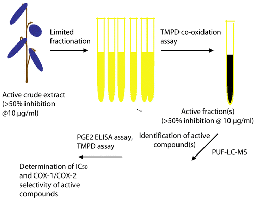
Figure 1.  Methods used for assessment of COX-2 activities in plant extracts using traditional enzyme assays and COX-2 PUF assay.

Figure 2. Schematic presentation of PUF-LC-MS assay.

Figure 2.  Schematic presentation of PUF-LC-MS assay.

Table 1. Minimum inhibitory concentration of a methanol extract of cranberry in two clinical E. coli strains. All experiments were performed in triplicate.

Table 2. Effect of various cranberry extracts on COX-2 activity in vitro. All experiments were performed in triplicate.

Figure 3. Structure of ursolic acid derivatives inhibiting COX-2 as determined using the COX-2 PUF-LC-MS assay.
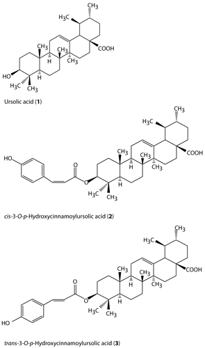
Figure 3.  Structure of ursolic acid derivatives inhibiting COX-2 as determined using the COX-2 PUF-LC-MS assay.

Figure 4. Cranberry extract (CRME) inhibits NF-κB-dependent transcriptional activities. CRME extract inhibited TNF-induced NF-κB activation in stably transfected human T lymphocytes 5.1 cells as assessed by a reporter gene assay. The luciferase activity was measured after 6 h and expressed as TNF induction = 100%. Values are means of ± SD of three independent experiments in triplicate.
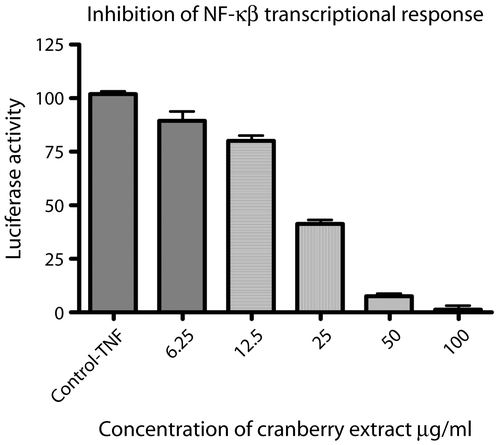
Figure 4.  Cranberry extract (CRME) inhibits NF-κB-dependent transcriptional activities. CRME extract inhibited TNF-induced NF-κB activation in stably transfected human T lymphocytes 5.1 cells as assessed by a reporter gene assay. The luciferase activity was measured after 6 h and expressed as TNF induction = 100%. Values are means of ± SD of three independent experiments in triplicate.

Figure 5. Cranberry extract inhibited cytokine release in human peripheral blood mononuclear leukocytes (PMNs). Cells were stimulated with E. coli LPS (25 ng/mL) for 6–16 h in the presence or absence of cranberry extract (0-100 μg/mL). Data are expressed as the percentage of cytokines accumulated in the supernatant of LPS-induced PMNs (100%) and represent the means ± SEM of at least three experiments in triplicate. P < 0.001 represents a significant difference compared to the values seen in the LPS-activated cells
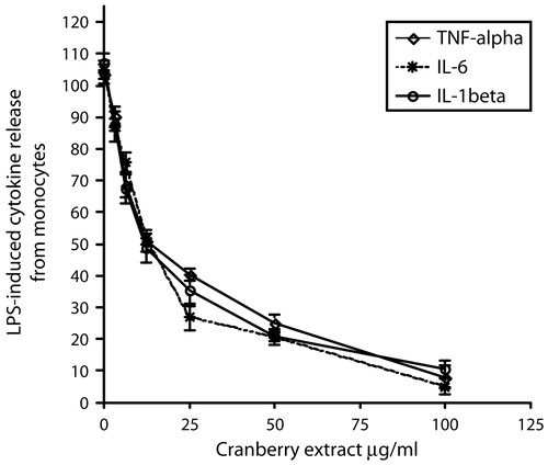
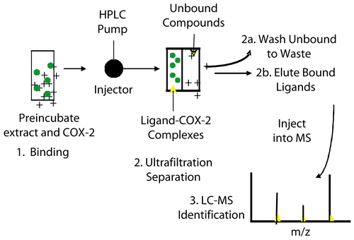
Figure 5.  Cranberry extract inhibited cytokine release in human peripheral blood mononuclear leukocytes (PMNs). Cells were stimulated with E. coli LPS (25 ng/mL) for 6–16 h in the presence or absence of cranberry extract (0-100 μg/mL). Data are expressed as the percentage of cytokines accumulated in the supernatant of LPS-induced PMNs (100%) and represent the means ± SEM of at least three experiments in triplicate. P < 0.001 represents a significant difference compared to the values seen in the LPS-activated cells

Reprints and Corporate Permissions

Please note: Selecting permissions does not provide access to the full text of the article, please see our help page How do I view content?

To request a reprint or corporate permissions for this article, please click on the relevant link below:

Academic Permissions

Please note: Selecting permissions does not provide access to the full text of the article, please see our help page How do I view content?

Obtain permissions instantly via Rightslink by clicking on the button below:

If you are unable to obtain permissions via Rightslink, please complete and submit this Permissions form. For more information, please visit our Permissions help page.