Figures & data
Table 1. Effect of leaf ethanolic extract of C. viscosum and PZQ on R. tetragona.
Figure 1. Light micrographs of proglottids of R. tetragona: (A) control showing uniform smooth outer tegument (ot), sub-tegument (st), muscular layer (ml); (B) C. viscosum and (C) PZQ-treated worms reveal invagination, cracks and infoldings in the outer tegument and muscular layer is disrupted. All scale bars 100 μm.
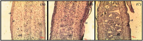
Figure 2. SEM of scolex of R tetragona: (A) Control showing scolex with wide suckers and smooth neck region, Scale bar 20 μm; (B) C. viscosum and (C) PZQ-treated parasites showing shrinkage in both scolex and suckers, Scale bars 10 μm and 20 μm, respectively.
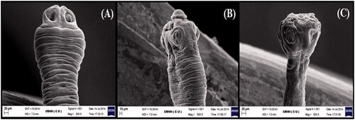
Figure 3. SEM of mature proglottids of R. tetragona: (A) control showing smooth velvety appearance; (B) treated worm with C. viscosum show vacuolization and no differentiation between each segment; (C) treated with PZQ show cracks and wrinkle formation in the tegument. All scale bars 10 μm.
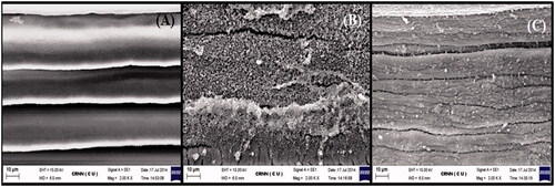
Figure 4. SEM of microtriches of R. tetragona: (A) control showing uniform and sharply arranged microtriches; (B) treated worms with C. viscosum and (C) PZQ reveal clumping of microtriches. All scale bars 1 μm.
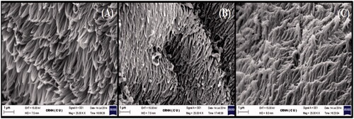
Figure 5. TEM of tegument of R. tetragona: (A) control showing an outer layer of dense microtriches (m) followed by thick syncytial (s) layer, muscle layers (ml) and parenchymal layer (p) and numerous secretory bodies (→); (B) worms treated with C. viscosum showing sloughed off microtriches, syncytial layer is exposed, muscle layer disoriented; (C) PZQ-treated worms showing clumping microtriches, syncytial layer degraded, muscle layer not distinct. All scale bars 1 μm.
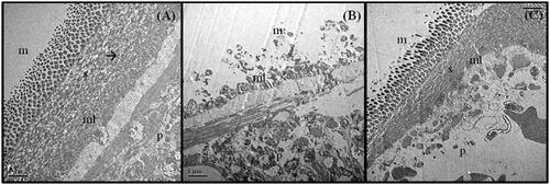
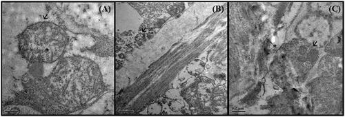
Figure 6. TEM of the nucleus of R tetragona: (A) control showing nucleus with distinct nuclear membrane and nucleolus and grannular cytoplasm; (B) treated worms with C. viscosum showing distinct nuclear membrane but with electron lucent matrix and distinct nucleolus; (C) PZQ-treated worms showing nucleus with dense cytoplasm. All scale bars 1 μm.
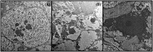
Figure 7. TEM of mitochondria (→) of R. tetragona: (A) control showing mitochondria with defined cristae and smooth membrane, scale bar 0.2 μm; (B) worms treated with C. viscosum and (C) PZQ showing fuzzy mitochondira; Scale bar 0.5 μm and 0.2 μm, respectively.