Figures & data
Figure 1. Study flowchart explaining the various groups of rats and treatments. ND: normal chow diet; HFD: high-fat diet; Dw: distilled water; Ac: acetic acid; PV: pomegranate vinegar; PPV: prickly pear vinegar; AV: apple vinegar.
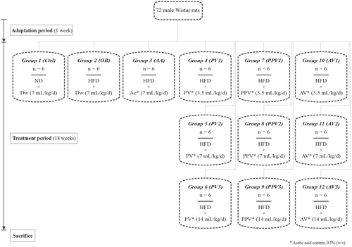
Table 1. Effect of fruit vinegars (FVs) on growth performance, food intake, and weight of heart and visceral adipose tissue (VAT).
Table 2. Effect of fruit vinegars (FVs) on lipids profile, leukocyte count, and plasma inflammatory and cardiac biomarkers.
Table 3. Effect of fruit vinegars (FVs) on plasma and visceral adipose tissue-derived (VAT) cytokines.
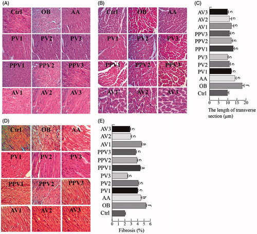
Figure 2. FVs attenuate cardiac histological abnormalities, hypertrophy and fibrosis in the hearts of HFD-fed rats. (A,B) Representative images for the hematoxylin-eosin (H&E) staining in the formalin-fixed myocardial tissues (400× amplification). (C) Quantitative data of myocyte cross-section length of 100 cells chosen from different visual scopes of three samples per group in myocardial transverse H&E staining are shown. (D) Representative images for the Masson staining in the formalin-fixed myocardial tissues (400× amplification). (E) Quantitative analysis of fibrotic area (Masson trichrome-stained area in light blue normalized to total myocardial area; n = 3 rats/group). Values are statistically different at: ap < .05, bp < .01, cp < .001, when compared with OB group values. Values are statistically different at: dp < .05, ep < .01, fp < .001, when compared with Ctrl group values.