Figures & data
Figure 1. GC chromatogram of essential oil of M. paniculata.
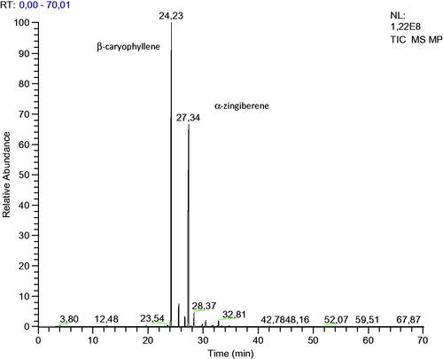
Table 1. Chemical composition of the essential oil of M. paniculata.
Table 2. Antioxidant activity of the M. paniculata essential oil and β-caryophyllene by the DPPH and ABTS methods.
Table 3. Minimum Inhibitory concentration (MIC), minimum bactericidal concentration (MBC), and minimum fungicidal concentration (MFC) of the essential oil of M. paniculata and β-caryophyllene.
Figure 2. Time–kill curve studies of M. paniculata essential oil and β-caryophyllene against S. aureus, E. coli, S. typhimurium and E. faecalis bacteria.
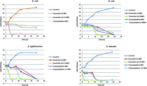
Figure 3. Time–kill curve studies of essential oil of M. paniculata and β-caryophyllene against the fungi A. niger, F. sollani, A. parasiticum and A. fumigatus.
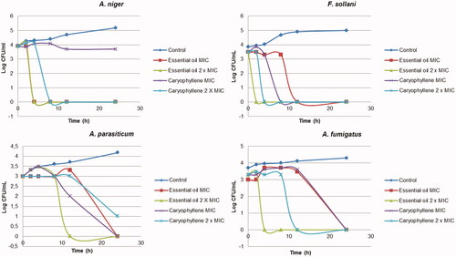
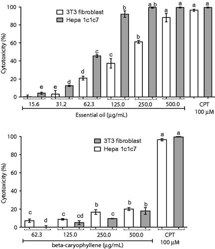
Figure 4. Cytotoxic activity of M. Paniculata essential oil and β-caryophyllene in 3T3 fibroblasts and Hepa 1c1c7, a human hepatoma cell line using the MTT assay. Data are presented as percentage of cell death from three independent experiments. Camptothecin was used as positive control. Different letters correspond to significant difference (p < 0.05); CPT: camptothecin.