
Figures & data
Table 1. Identification and antagonistic activity of endophytic fungi isolated from the leaves of Markhamia tomentosa.
Figure 1. The morphology (colony appearance, fruiting structures and spores) of identified endophytic fungi isolated from the leaves of Markhamia tomentosa.
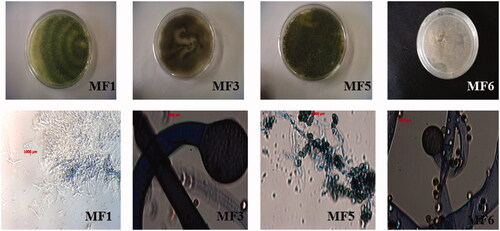
Figure 2. Dual culture bioassay of endophytic fungi of Markhamia tomentosa against Fusarium oxysporum (F. o; ITCC 3636) and Sclerotinia sclerotorium (S. s; ITCC 6323).
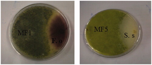
Table 2. Yield of extracts obtained from endophytic fungi isolated from Markhamia tomentosa.
Table 3. Antiproliferative, antifungal and antifeedant activity of fungal crude extracts and fractions.