Figures & data
Table 1. Quantification of total phenolics and tannins content present in the ethanol leaf and bark extracts of S. australis.
Figure 1. Representative chromatogram of the ethanol leaf extract of S. australis (under conditions described in LC-ESI-MS/MS analyses).
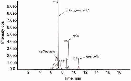
Table 2. In vitro antioxidant activity of ethanol leaf and bark extract of S. australis determined by DPPH radical scavenging activity, ferric reducing antioxidant power (FRAP) and nitric oxide radical scavenging assay.
Table 3. Antimicrobial activity of the ethanol leaf and bark extracts of S. australis expressed as Minimum Inhibitory Concentration (MIC) in μg/mL.
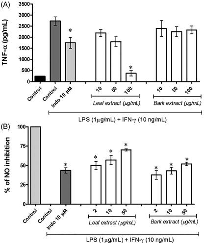
Figure 2. S. australis reduce nitric oxide (NO) and TNF-α concentration in vitro. RAW 264.7 macrophage were exposed with or without ethanol leaf and barks extracts of S. australis for 2 h and then stimulated with LPS/IFN-γ. (A) TNF-α and (B) NO production was measured 24 h later using ELISA Kit and the Griess reagent, respectively, as described in the “Materials and methods” section. Results are expressed as mean ± SD of three independent experiments. *Significant at p < 0.05 compared to control LPS/IFN-γ-induced cells.