Figures & data
Table 1. Effects of Vacha rhizome extract on body and lymphoid organ weight of rats.
Table 2. Effects of Vacha rhizome extract on adrenal 3β-HSDH activity and weight of the adrenal gland of rats.
Table 3. Effects of Vacha rhizome extract on total and differential counts of leukocytes of rats.
Table 4. Effects of Vacha rhizome extract on circulating immune complexes and total immunoglobulins contents of rats.
Table 5. Effects of Vacha rhizome extract on bone marrow stem cells of rats.
Table 6. Effects of Vacha rhizome extract on lymphocyte counts in different lymphoid organs of rats.
Figure 1. (a–e) Photomicrographs of splenocytes stained with acridine orange and ethidium bromide (450 nm and 530 nm). Note the presence of more number of healthy cells (N) in control (a), vehicle control (b), unstressed + Vacha extract treated rats (c) and stress + Vacha extract treated rats (e), and more number of apoptotic cells (A) in stressed (d) rats. 200×. A: apoptotic cells; N: normal, healthy cells.

Table 7. Effects of Vacha rhizome extract on apoptosis of cells in lymphoid organs of rats.
Figure 2. (a–e) Photomicrographs of the cross sections of the spleen showing the island of white pulp. Note the shrinkage of island of the white pulp region and germinal center in stressed rats (d) compared to control (a), vehicle control (b), unstressed + Vacha extract (c) and stress + Vacha extract (e) treated rats. 200× (H&E). GC: germinal center; CA: central artery; L: lymphocytes.
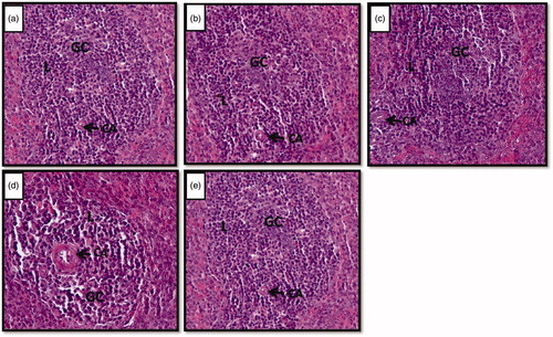
Table 8. Effects of Vacha rhizome extract on number of islands of white pulps in spleen of rats.
Figure 3. (a–e) Photomicrographs of the cross sections of thymus gland. Note the reduction of cortex region and development of connective tissue and fat cells in stressed (d) rats compared to control (a), vehicle control (b), unstressed + Vacha extract (c) and stress + Vacha extract (e) treated rats. 200× (H&E). C: connective tissue fibers; F: fat cells.
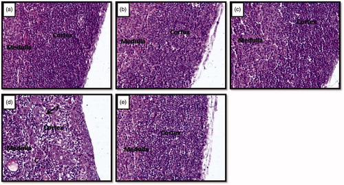
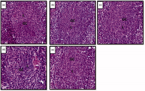
Figure 4. (a–e) Photomicrographs of cross sections of axillary lymph node showing lymphoid follicles. Note the shrinkage of germinal centers in stressed (d) rat compared to control (a), vehicle control (b), unstressed + Vacha extract (c) and stress + Vacha extract (e) treated rats. 200× H&E. GC: germinal centre; L: B lymphocytes.