
Figures & data
Table 1. Composition of modified Zarrouk medium.
Figure 1. Cell culture insert model.
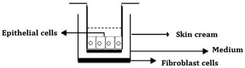
Figure 2. Wound creating apparatus and making a scratch in 24-well plate.
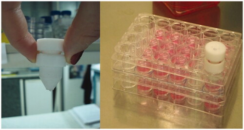
Figure 3. In vitro cytotoxicity of S. platensis extracts on HS2 keratinocyte cell line.
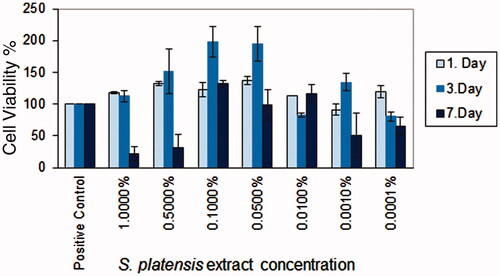
Figure 4. Morphological observation of the wounded edge and centre of HS2 cell cultures. (a,b) Negative cell control (4×, 10×); (c,d) cream control (4×, 10×); (e,f) cream with 0.5% Spirulina platensis extract (4×, 10×); (g,h) cream with 1.125% S. platensis extract (4×, 10×) cell migration and wound healing for 5 d.
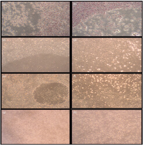
Table 2 Wound closure effects of negative cell control, skin cream and crude extract incorporated skin cream.
Figure 5. Immunohistochemical staining results of HS2 cell cultures. (a,b) Negative cell control (5 d, 10 d); (c,d) cream with 0.5% Spirulina platensis extract (5 d, 10 d); (e,f) cream with 1.125% S. platensis extract (5 d, 10 d); (g,h) cream control (5 d, 10 d) and (i,j) IHC control staining (5 d, 10 d).

Figure 6. Micronucleus formation during incubation with skin cream extracts.
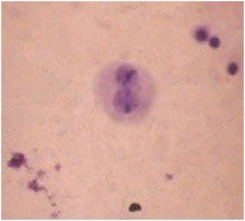
Table 3. Distribution of micronuclei (MN) in the binucleated (BN) cells scored, total number of MN, mean ± SD.