Figures & data
Figure 1. (a) After acclimatization in individual cages for two weeks, the rats were conditioned to take water only once a day for 30 min at a specified time on each day (water deprivation schedule of 23.5 h, ‘one bottle regime’). On day 0, the conditioned rats were selected on the basis of preference for intake of saccharin by using ‘two bottle regime’ where animals were given both water and saccharin simultaneously for 30 min only. Rats showing ≥50% intake of saccharin of the total fluid intake were selected. Selected rats were randomized and divided into three groups. Group I: untreated control (UC), group II: 2 Gy, group III: SBL-1 + 2 Gy. CTA was assessed in terms of saccharin preference ratio (SPR); {SPR (%) = [saccharin intake/(water intake + saccharin intake)] × 100}, decrease in SPR indicated acquisition of CTA. (b) It shows SPR (%) change in different groups. Data are presented as mean ± SD of six rats after normalization with respect to UC. The SPR was 100% for UC. *statistically significant in comparison with UC at p < 0.05.
![Figure 1. (a) After acclimatization in individual cages for two weeks, the rats were conditioned to take water only once a day for 30 min at a specified time on each day (water deprivation schedule of 23.5 h, ‘one bottle regime’). On day 0, the conditioned rats were selected on the basis of preference for intake of saccharin by using ‘two bottle regime’ where animals were given both water and saccharin simultaneously for 30 min only. Rats showing ≥50% intake of saccharin of the total fluid intake were selected. Selected rats were randomized and divided into three groups. Group I: untreated control (UC), group II: 2 Gy, group III: SBL-1 + 2 Gy. CTA was assessed in terms of saccharin preference ratio (SPR); {SPR (%) = [saccharin intake/(water intake + saccharin intake)] × 100}, decrease in SPR indicated acquisition of CTA. (b) It shows SPR (%) change in different groups. Data are presented as mean ± SD of six rats after normalization with respect to UC. The SPR was 100% for UC. *statistically significant in comparison with UC at p < 0.05.](/cms/asset/4b3cd01f-abd9-44ac-8af0-2da835db4304/iphb_a_1331365_f0001_b.jpg)
Figure 2. Modifying effect of Hippophae leaf extract (SBL-1) on radiation-induced changes in the levels of (a) dopamine, (b) norepinephrine and (c) epinephrine in brain and plasma of rats. Data are presented as mean ± SD of six rats in each group. *significantly different in comparison with untreated control (UC, group I) at p < 0.05 and #significantly different in comparison with 2 Gy (group II) at p < 0.05.
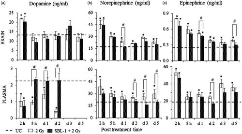
Figure 3. Radiation-induced changes and their modification by Hippophae leaf extract (SBL-1) on the levels of (a) serotonin and (b) acetylcholinesterase (AChE) in brain tissue of rats. Data are presented as mean ± SD of six rats in each group. *significantly different in comparison with untreated control (UC, group I) at p < 0.05 and #significantly different in comparison with 2 Gy (group II) at p < 0.05.
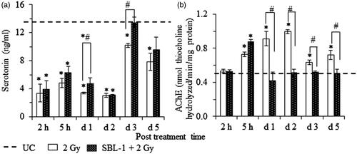
Table 1. Effect of Hippophae leaf extract (SBL-1) on radiation-induced changes in the levels of superoxide dismutase (SOD), catalase (CAT) and reduced glutathione (GSH) in brain and blood of rats.
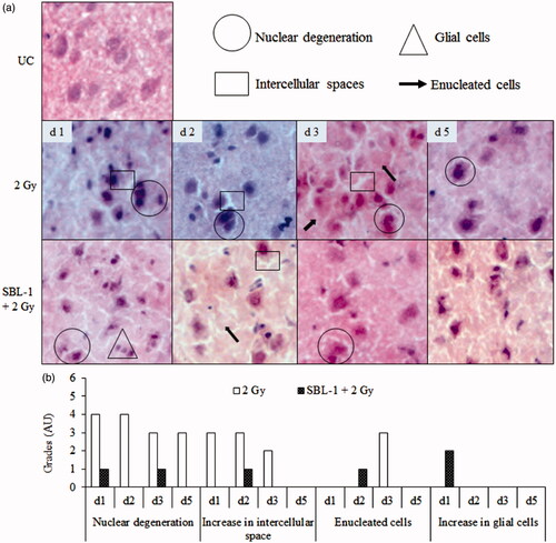
Figure 4. Effect of Hippophae leaf extract (SBL-1) on radiation-induced histological changes in cerebral cortex of rats. (a) Microscopic observations of haematoxylin- and eosin-stained tissue sections are presented at 200 × magnification. (b) Changes with respect to untreated control (UC, group I) are graded on number scale with 0 as minimum and 6 as maximum using arbitrary units (AU).
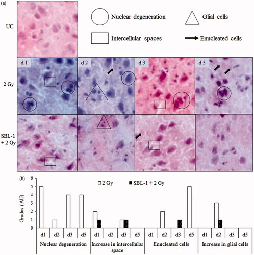
Figure 5. Effect of Hippophae leaf extract (SBL-1) on radiation-induced histological changes in hippocampus (Hipp) of rat brain. (a) Microscopic observations of haematoxylin- and eosin-stained tissue sections are presented at 50 × and 200 × magnifications. (b) Changes with respect to untreated control (UC, group I) are graded on number scale with 0 as minimum and 6 as maximum using arbitrary units (AU).
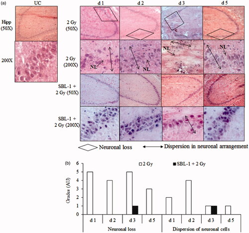
Figure 6. Effect of Hippophae leaf extract (SBL-1) on radiation-induced histological changes in amygdala of rats. (a) Microscopic observations of haematoxylin- and eosin-stained tissue sections are presented at 200 × magnification. (b) Changes with respect to untreated control (UC, group I) are graded on number scale with 0 as minimum and 6 as maximum using arbitrary units (AU).