Figures & data
Figure 1. Effects of AEDKC on ovalbumin-induced active systemic anaphylaxis. The induction of systemic anaphylaxis and oral administration of drugs, including AEDKC and Dexa, are described in the Materials and methods section. Blood was obtained from the abdominal artery of each mouse and measurements of serum histamine, total IgE, OVA-specific IgE, and IL-4 were taken. (A) Rectal temperature was measured every 10 min for 1 h. (B) Rectal temperature of the mice at 50 min. (C–F) Serum levels of histamine, total IgE, OVA-specific IgE, and IL-4. Graph data represent the mean ± SD (n = 5/group) of two independent experiments. *p < 0.05 compared with the OVA-challenged group. Dexa: dexamethasone.
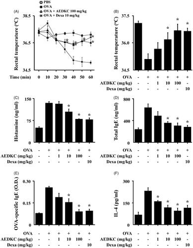
Figure 2. Effects of AEDKC on IgE-mediated passive cutaneous anaphylaxis. Mouse ear skin (n = 5/group) was sensitized with an intradermal injection of anti-DNP IgE (0.5 μg/site) for 48 h. AEDKC and Dexa were orally administered 2 h before the intravenous injection of DNP-HSA (1 mg/mouse) and 4% Evans blue (1:1) mixture. Thirty minutes later, the ears were collected to measure pigmentation. The dye was extracted as described in the Materials and methods section and detected using a spectrophotometer. (A) Representative photographic images of ears. (B) Graph data represent the mean ± SD (n = 5/group) of two independent experiments. *p < 0.05 compared with the DNP-HSA-challenged group. Dexa: dexamethasone.
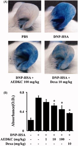
Figure 3. Effects of AEDKC on intracellular calcium and mast cell degranulation. (A) RBL-2H3 cells (6 × 10⁴/well) were pretreated with or without AEDKC for 12 h and then incubated with 1 mg/mL MTT for 2 h. The absorbance intensity was measured using a spectrophotometer. (B,C) RBL-2H3 cells (5 × 10⁵/well) and RPMCs (2 × 10⁴/well) were sensitized with anti-DNP IgE (50 ng/mL). After overnight incubation, the cells were pretreated with or without drugs, including AEDKC and Dexa, for 1 h, and then challenged with DNP-HSA (100 ng/mL). (D) The level of β-hexosaminidase was measured using β-hexosaminidase substrate buffer. RBL-2H3 cells (6 × 10⁴/well) were sensitized with anti-DNP IgE. (E) After overnight anti-DNP IgE incubation, cells were incubated with Fluo-3/AM for 1 h, treated with or without AEDKC for 1 h, and then challenged with DNP-HSA. Intracellular calcium was detected using a fluorescent plate reader. BAPTA-AM, a calcium chelator, was used as the positive control. Graph data represent the mean ± SD of three independent experiments. *p < 0.05 compared with the DNP-HSA-challenged group. Dexa: dexamethasone.
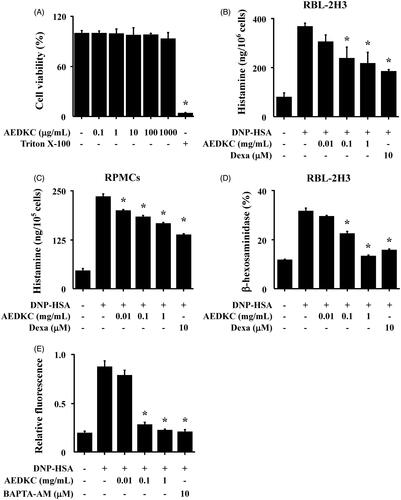
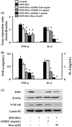
Figure 4. Effects of AEDKC on inflammatory cytokines and NF-κB activation in mast cells. RBL-2H3 cells (5 × 10⁵/well) were sensitized with anti-DNP IgE (50 ng/mL). After overnight incubation, the cells were pretreated with or without drugs, including AEDKC and Dexa, for 1 h, and then challenged with DNP-HSA (100 ng/mL). (A) The gene expression of inflammatory cytokines was determined by qPCR. (B) The secretion of inflammatory cytokines was measured by ELISA. Graph data represent the mean ± SD of three independent experiments. (C) NF-κB activation was assayed by Western blot (N: nuclear). β-actin and lamin B were used as loading controls. The bands are representative of three independent experiments. *p < 0.05 compared with the DNP-HSA-challenged group. Dexa: dexamethasone.