Figures & data
Figure 1. GC-MS chromatogram of the essential oil of Zataria multiflora.
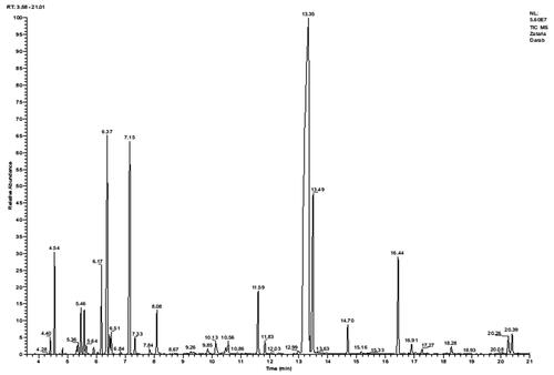
Table 1. Chemical constituents of Zataria multiflora essential oil.
Figure 2. The effects of topical administration of ZMEO on (B) wound contraction rate and (C) tissue bacterial count (CFU/g) in different days. Superscripts (a–d) indicate significant differences in same day at p < 0.05.
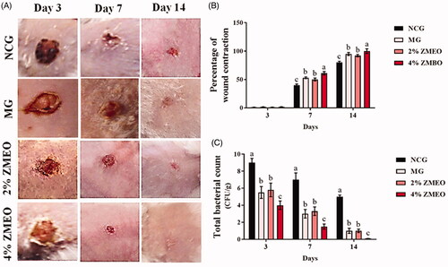
Figure 3. Immunohistochemical staining for angiogenesis:; (A) NCG group, (B) MG group, (C and D) 2 and 4% ZMEO-treated groups. See elevated angiogenesis in ZMEO-treated group (arrows) on day 7 after wound induction. CD 31 staining, 400×.
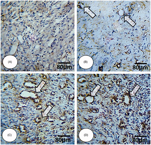
Figure 4. Cross section from wound area: (A) NCG group, (B) MG group, (C and D) 2 and 4% ZMEO-treated groups. Note well-formed collagen deposition in cross sections from ZMEO-treated animals on day 7 after wound induction (first row), which is significantly increased on day 14 after injury (second row). The third row represents the software analyze for collagen intensity. The cross sections from animals in ZMEO-treated groups exhibited condense collagen deposition versus NCG and MG groups. Masson-trichrome staining and fluorescent staining for collagen, 400× magnification.
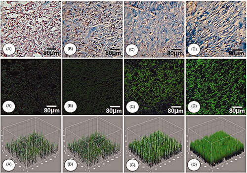
Figure 5. Cross section from wound area: (A) NCG group, (B) MG group, (C and D) 2 and 4% ZMEO-treated groups. See well re-epithelialization in ZMEO-treated animals. The re-epithelialization initiated on day 7 after wound induction in ZMEO-treated animals. However, the cross sections from NCG and MG groups are not representing epithelialization. Note well-organized dermis and complete epithelialization with well-formed papillae in ZMEO-treated animals in comparison to NCG and MG groups. Masson-trichrome staining, 100×.
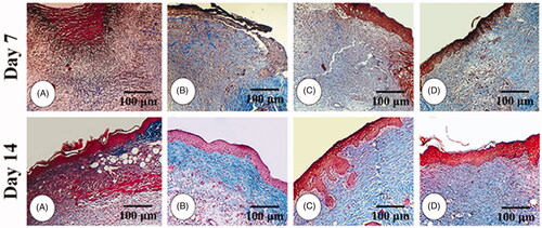
Table 2. The effects of ZMEO on histopathological parameters in different days.
Figure 6. The effects of topical application of ZMEO on gene expression. Superscripts (a–d) indicate significant differences in same day at p < 0.05.
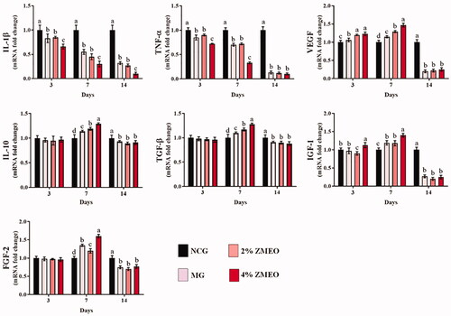
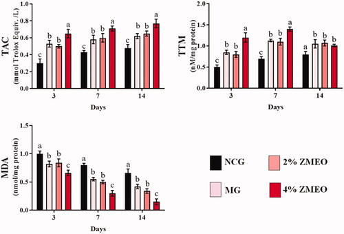
Figure 7. The effects of topical application of ZMEO on antioxidant properties. Superscripts (a–d) indicate significant differences in same day at p < 0.05.