Figures & data
Figure 1. Effect of berberine on cardiac hemodynamics in a rat model of sepsis. (A) LVSP (mmHg) at 6, 24, and 48 h; (B) LVEDP (mmHg) at 6, 24, and 48 h; (C) +dp/dt max (mmHg) at 6, 24, and 48 h; (D) –dp/dt max (mmHg) at 6, 24, and 48 h. (E) Levels of cTnT measured by ELISA at 6, 24, and 48 h. *p < 0.05 indicates significance in comparison with the Con group; △p < 0.05 indicates significance in comparison with the LPS group.
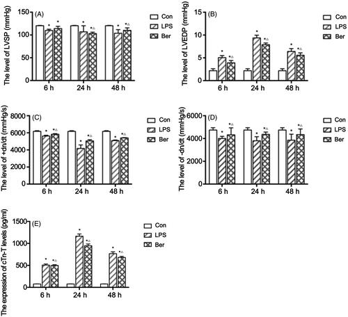
Figure 2. Histological changes of the myocardial tissue assessed by H&E staining. (A, B, C) Histopathological features of the myocardial tissue in the Con group at 6, 24, and 48 h. (D, E, F) Cardiomyocytes were swollen and enlarged, with infiltrated lymphocytes emerging in the LPS group at 6, 24, and 48 h. (G, H, I) Histopathological features of the myocardial tissue in the Ber group at 6, 24, and 48 h. Magnification, 200X.

Figure 3. Effects of berberine on key effectors of the TLR4/NF-κB signalling pathway. (A) TLR4 protein expression in the myocardial tissue and quantitation. (B) Nuclear p65 protein expression in the rat sepsis cardiomyopathy model measured by ELISA. Effects of berberine on TNFα (C) and IL 1β (D) levels in the myocardial tissue. *p < 0.001 indicates significance in comparison with the Con group. △p < 0.001 indicates significance in comparison with the LPS group.
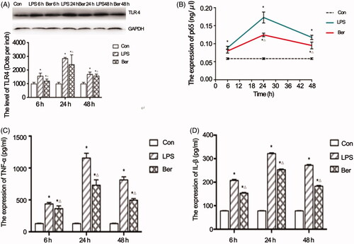
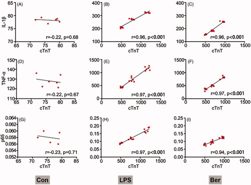
Figure 4. Correlation between cTnT and NF-κB signalling. Associations of cTnT amounts with IL-1β, TNFα and p65 levels in the Con (A, B, C), LPS (D, E, F) and Ber (G, H, I) groups, assessed by Pearson correlation analysis r correlation coefficient. p < 0.05 indicates a valid correlation.