
Figures & data
Figure 1. The cytotoxicity of Phy in breast cancer cells. (A) Phy suppressed the cell viability of MCF-7 cells measured by MTT assay after 24 h exposure. (B) Phy enhanced the release of LDH in MCF-7 cells. Twenty-four hour Phy incubation enhanced the activities of (C) caspase-3, (D) caspase-8, and (E) caspase-9 in MCF-7 cells. (F) Phy enhanced the apoptosis rate of MCF-7 cells after 24 h exposure. Data are expressed as percentages relative to the corresponding control cells and as mean ± S.D. (n = 6). *p<0.05, **p<0.01, and ***p<0.001 versus control cells.
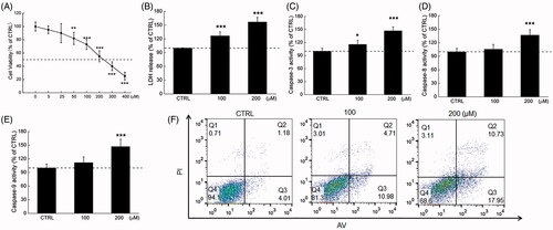
Figure 2. Phy caused mitochondrial dysfunction in breast cancer cells. Phy (A) upregulated ROS production and (B) decreased the MMP (20× magnification; scale bar: 100 μm) in MCF-7 cells after 24 h treatment. (C) Phy distinctly reduced the expression levels of Bcl-2 and Bcl-xL, and enhanced the expression levels of Bax in MCF-7 cells. Quantitative protein expression data were normalised to GAPDH levels in the corresponding samples. Data are expressed as percentages relative to the corresponding control cells and mean ± S.D. (n = 6). **p<0.01 and ***p<0.001 versus control cells.
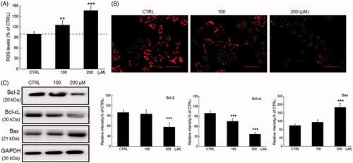
Figure 3. Phy suppressed MCF-7-xenograft tumour growth in BALB/c nude mice. MCF-7-xenograft BALB/c nude mice model were treated with Phy at 30 mg/kg every other day for 14 days. (A) Tumour-bearing nude mice. (B) Tumour tissue specimens. (C) Tumour sizes of MCF-7-xenografted nude mice in the control and Phy-treated groups. Tumour sizes are expressed as mean ± S.D. (n = 3). *p<0.05 versus control group. (D) Mean (± S.D.) body weight of the control and Phy-treated mice (n = 3). (E) Phy enhanced the expression levels of Bax and cleaved caspase-3, and reduced the expression levels of Bcl-2, Bcl-xL, Nrf2 and its downstream proteins. Quantitative protein expression data were normalised to the corresponding GAPDH levels, and the average fold changes in band intensity are marked (n = 3). Haematoxylin and eosin staining of liver (F) and spleen (G) tissues from nude mice.
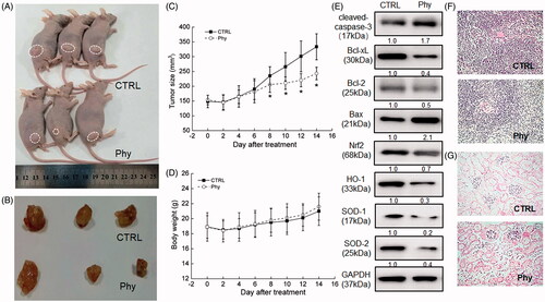
Figure 4. Phy suppressed MCF-7-xenograft tumour growth in BALB/c mice. The BALB/c mouse model was treated with Phy at 30 mg/kg every other day for 28 days. (A) Tumour-bearing nude mice. (B) Tumour tissue specimens. (C) Tumour sizes of MCF-7-xenografted mice in the control and Phy-treated groups. Tumour sizes are expressed as mean ± S.D. (n = 3). ***p < 0.001 versus control group. (D) Mean (± S.D.) body weight of the control and Phy-treated mice (n = 3). (E) Phy enhanced the phosphorylation of IκBα and NF-κB, and reduced the expression levels of Nrf2 and its downstream proteins. Quantitative protein expression data were normalized to the corresponding GAPDH levels, and the average fold changes in band intensity are marked (n = 3). Haematoxylin and eosin staining of liver (F) and spleen (G) tissues from nude mice.
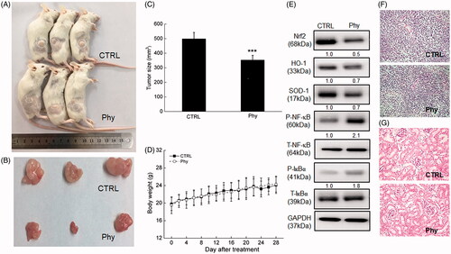
Table 1. The effects of Phy on the immune factors in serum of MCF-7-xenografted BALB/C mice.
Table 2. The effects of Phy on oxidative factors of serum in tumour- xenografted BALB/c mice.