Figures & data
Figure 1. Chemical structure of myrtenol.
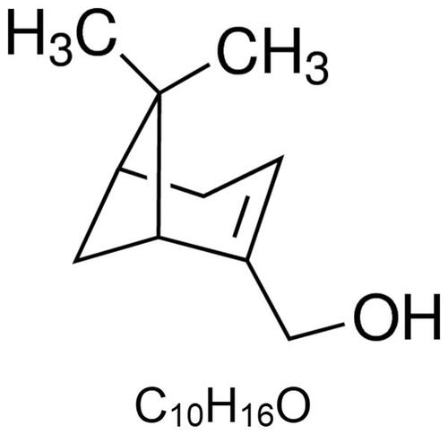
Figure 2. Myrtenol improved neurological function and cerebral infarction of MCAO rats. (A) The neurological score of rats in each group at 24 h after administration (n = 15). (B) The frequency of mistakes by rats in each group was detected by diving platform experiment (n = 15). (C) The times of entries arm of rats in each group was detected by Y-maze testing (n = 15). (D) The brain water content of rats in each group (n = 5). (E) Effect of myrtenol on the cerebral infarct volume of rats in each group was measured by TTC staining (n = 4). (F-H) The expression of BDNF and NGF in brain tissues of rats in each group was detected by western blot (n = 6). Data were presented as the mean ± SD of at least three repeated experiments. ##p < 0.01, compared with the sham group; *p < 0.05, **p < 0.01, compared with the MCAO group. Except for the sham group, all rats in other groups were constructed for cerebral I/R injury by MCAO. Sham group and MCAO group, given saline after cerebral I/R surgery. MCAO + Myr groups, administered 10, 30, or 50 mg/kg myrtenol after cerebral I/R surgery, respectively. All rats were treated once daily for seven consecutive days.
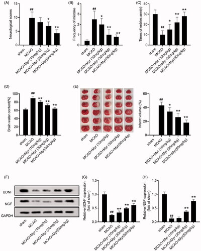
Figure 3. Myrtenol improved hippocampus damage and reduced cell apoptosis in MCAO rats. (A) Histological damage and apoptosis of hippocampus were analysed using HE staining and TUNEL assay (n = 6). (B–D) The expression of apoptosis marker protein, Bcl-2 and Bax, in hippocampus of rats in each group was detected by western blot (n = 6). (E) The caspase-3 activity in hippocampus of rats in each group (n = 6). Data were presented as the mean ± SD of at least three repeated experiments. ##p < 0.01, compared with the sham group; *p < 0.05, **p < 0.01, compared with the MCAO group.
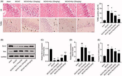
Figure 4. Mytenol promoted angiogenesis in MCAO rats. (A–B) VEGF expression in ischaemic penumbra were determined by IHC staining (n = 6). (C–E) The relative expression of VEGF and FGF2 protein was detected by western blot (n = 6). Data were presented as the mean ± SD of at least three repeated experiments. ##p < 0.01, compared with the sham group; *p < 0.05, **p < 0.01, compared with the MCAO group.
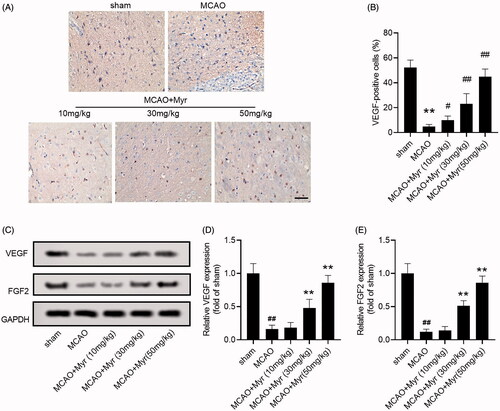
Figure 5. Mytenol activated ERK1/2 signalling pathway in MCAO rats. (A) MEK1/2, p-MEK1/2, ERK1/2, p-ERK1/2 expression in brain tissues were detected by western blot. (B–C) The relative expression ratio of p-MEK1/2/MEK1/2, p-ERK1/2/ERK1/2. (D) MEK1/2, p-MEK1/2, ERK1/2, p-ERK1/2 expression in brain tissues were detected by western blot. (E-F): MEK1/2, p-MEK1/2, ERK1/2, p-ERK1/2 expression in brain tissues were detected by western blot. n = 6. Data were presented as the mean ± SD of at least three repeated experiments. ##p < 0.01, compared with the sham group; *p < 0.05, **p < 0.01, compared with the MCAO group.
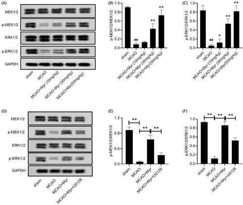
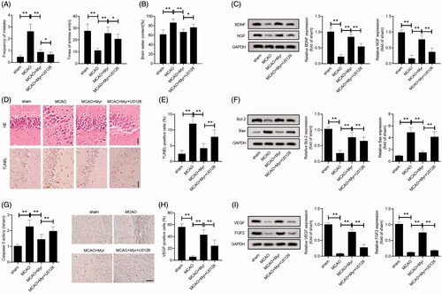
Figure 6. U0126 reversed the effect of myrtenol on the improvement of brain damage and angiogenesis in MCAO rats. (A) Frequency of mistakes, times of entries arm of rat in each group were detected by diving platform experiment, Y-maze testing (n = 15). (B) Brain oedema was assessed by brain water content (n = 5). (C) Relative expression of BDNG and NGF in brain tissues was detected by western blot (n = 6). (D–E) Histological damage and apoptosis of hippocampus were analysed using HE staining and TUNEL assay (n = 6). (F) Relative expression of Bcl-2, Bax was detected by western blot (n = 6). (G) The caspase-3 activity. (H) IHC staining showed the VEGF positive cells (n = 6). (I) Relative expression of VEGF and FGF2 was detected by western blot (n = 6). Data were presented as the mean ± SD of at least three repeated experiments. **p < 0.01. Except for sham group, all rats in other groups were constructed for cerebral I/R injury by MCAO. sham group and MCAO group, given with saline after cerebral I/R surgery. MCAO + Myr group, administered 50 mg/kg myrtenol after cerebral I/R surgery; MCAO + Myr + U0126 group, injected with U0126 at 30 min prior to myrtenol treatment. All rats were administered once daily for 7 consecutive days.