Figures & data
Figure 1. SQBD promotes apoptosis in CGM1 cells. CGM1 cells were treated with 10%-medicated serum or 10%-blank serum for 24 or 48 h. 10% FBS-treated CGM1 cells served as NC. (A and B) Flow cytometry was performed to detect apoptosis of the CGM1 cells after 24 or 48 h serum treatment. (C) The Caspase-Glo® 3/7 assay was performed to detect the activity of caspase 3/7 in the CGM1 cells. (D) WB was performed to estimate the expression of p53 in the CGM1 cells. (**p < 0.01 vs. 10%-blank serum group).
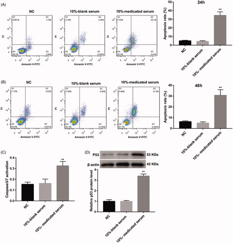
Table 1. Effect of the drug-contained serum on cell viability of CGM1 cells.
Figure 2. SQBD alters EBV latency protein expression in CGM1 cells. CGM1 cells were treated with 10%-medicated serum or 10%-blank serum. 10% FBS-treated CGM1 cells served as NC. (A) WB was performed to detect the expression of EBV latency proteins (EBNA2, LMP1, EBNA3A and EBNA3C) in the CGM1 cells. (B) WB was performed to assess the expression of p65, p-p65, p52, p100, c-MYC and cyclin E in the CGM1 cells. (*p < 0.05, **p < 0.01 vs. 10%-blank serum group).
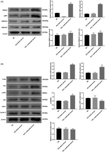
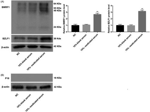
Figure 3. SQBD inhibits lytic EBV replication in CGM1 cells. CGM1 cells were treated with 10%-medicated serum or 10%-blank serum. 10% FBS-treated CGM1 cells served as NC. (A) WB was performed to explore the expression of immediate early lytic protein BZLF1 and the early lytic protein BMRF1 in the CGM1 cells. (B) WB was performed to assess the expression of p18 in the CGM1 cells. (**p < 0.01 vs. 10%-blank serum group).