Figures & data
Table 1. Composition of the modified Seonghyangjeongki-san (SHJKSm).
Figure 1. Schematic diagram of the process used to establish the middle cerebral artery occlusion (MCAO) model. The methanol fraction of the modified Seonghyangjeongki-san water extract (SHJKSmex) was administered for 3 consecutive days as pre-treatment before 2 h of MCAO induction. Mice were sacrificed 24 h after the initiation of MCAO (24 h post-MCAO). During the entire ischaemic period, the relative cerebral blood flow (rCBF) was monitored using laser Doppler flowmetry. LCCA: left common carotid artery; MCA: middle cerebral artery.
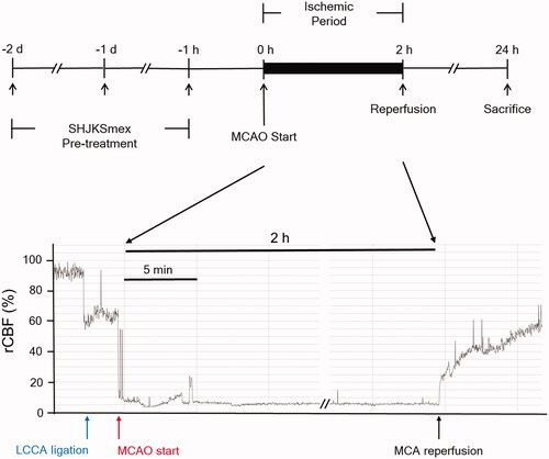
Figure 2. Relative cerebral blood flow (rCBF) in all groups (n = 6 per group). (A), Sham-operated normal group; (B), MCAO control group; (C), (D), and (E), SHJKSmex pre-treatment (30, 100, and 300 mg/kg SHJKSmex, respectively) groups. CCAL: common carotid artery; MCAO: middle cerebral artery occlusion; PM: pre-MCAO; RP: reperfusion; SHJKSmex: methanol fraction of the modified Seonghyangjeongki-san water extract.
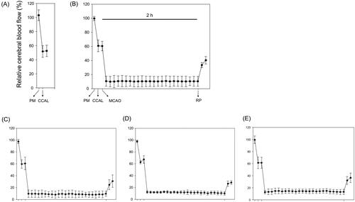
Figure 3. Influence of middle cerebral artery occlusion (MCAO)-induced brain injury and the effect of pre-treatment with the methanol fraction of the modified Seonghyangjeongki-san water extract (SHJKSmex) on body weight changes (A) and physiological parameters (B). Mice were weighed daily during the 4-day experimental period. Serum samples were obtained, and the concentrations of Na+, K+, and Cl− were measured. Results are presented as the mean ± SD.
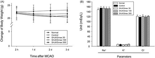
Figure 4. Representative group images and total infarct lesion volumes detected via TTC staining after 2 h of middle cerebral artery occlusion (MCAO) and the effects of pre-treatment with the methanol fraction of the modified Seonghyangjeongki-san water extract (SHJKSmex) on the total infarct volume (A) and neurological deficit scores (B) in each group. The harvested brain slices were stained with TTC to measure the infarct volumes. Ischaemic regions were identified as pale regions in the coronal slices. SHJKSmex pre-treatment significantly decreased the infarct volumes at 24 h after MCAO. SHJKSmex pre-treatment did not improve the neuronal deficit scores at 24 h after MCAO. The results are presented as the mean ± SD. ###p < 0.001 vs. normal group, *p < 0.05, ***p < 0.001 vs. MCAO control group; n = 6 in each group. TTC: 2,3,5-triphenyl-tetrazolium chloride.
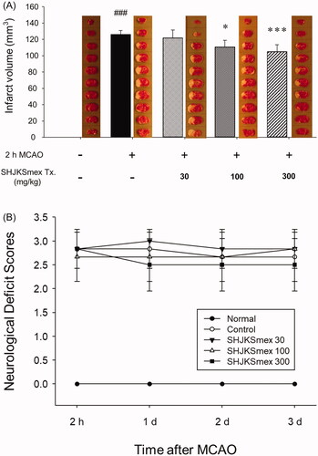
Figure 5. Influence of middle cerebral artery occlusion (MCAO)-induced brain injury and the effects of pre-treatment with the methanol fraction of the modified Seonghyangjeongki-san water extract (SHJKSmex) on brain edoema indices (A), brain water content (B), and aquaporin-4 (AQP-4) protein expression (C) in the brains of mice with MCAO. Pre-treatment with SHJKSmex caused no significant changes in the brain edoema index. Pre-treatment with 300 mg/kg SHJKSmex significantly suppressed the whole brain water content. SHJKSmex pre-treatment had no suppressive potential on AQP-4 overexpression; however, the values were considered to display a decreasing tendency. Western blots and quantitative analysis of AQP-4 protein expression in the brain tissue revealed similar results to the brain edoema index and brain water content findings. Results are presented as the mean ± SD. #p < 0.05, ###p < 0.001 vs. normal group, *p < 0.05 vs. control group; n = 6 per group.
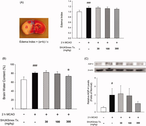
Figure 6. Effects of pre-treatment with the methanol fraction of the modified Seonghyangjeongki-san water extract (SHJKSmex) on reactive oxygen species (ROS) (A) and malondialdehyde (MDA) (B) levels in the brains of mice with middle cerebral artery occlusion (MCAO). Pre-treatment with 300 mg/kg SHJKSmex significantly lowered ROS and MDA levels. Results are presented as the mean ± SD. ###p < 0.001 vs. normal group, **p < 0.01 vs. control group; n = 6 in each group.
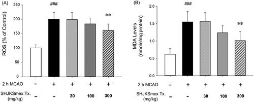
Figure 7. Effects of pre-treatment with the methanol fraction of the modified Seonghyangjeongki-san water extract (SHJKSmex) on inducible nitric oxide synthase (iNOS) (A), interleukin (IL)-1β (B), and tumour necrosis factor (TNF)-α levels (C) in the brains of mice with middle cerebral artery occlusion (MCAO). SHJKSmex pre-treatment significantly decreased iNOS levels in the mouse model of ischaemic brain stroke. Representative western blots and quantitative analysis of iNOS expression demonstrate the effect of SHJKSmex on iNOS expression in the brain tissue. IL-1β and TNF-α levels were measured using a commercially available ELISA kit. Results are presented as the mean ± SD. ###p < 0.001 vs. normal group, *p < 0.05, **p < 0.01 vs. control group; n = 6 in each group.
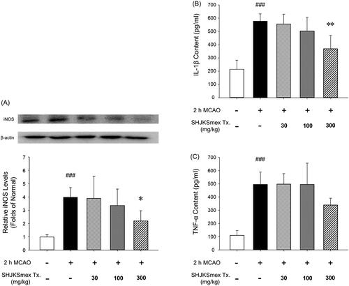
Figure 8. Effects of pre-treatment with the methanol fraction of the modified Seonghyangjeongki-san water extract (SHJKSmex) on the Bcl-2/Bax ratio (A) and caspase-8 (B) and caspase-9 (C) protein expression in the brains of mice with middle cerebral artery occlusion (MCAO). SHJKSmex pre-treatment significantly increased the Bcl-2/Bax ratio in the mouse model of ischaemic brain stroke. Representative western blots and quantitative analysis of Bcl-2 and Bax protein expression (A) revealed the upregulating potential of SHJKSmex on the Bcl-2/Bax ratio in the brain tissue. Representative western blots and quantitative analysis of caspase-8 and caspase-9 protein expression (B) revealed the potential of SHJKSmex to downregulate caspase-8 and caspase-9 protein expression in the brain tissue. Results are presented as the mean ± SD. #p < 0.05, ##p < 0.01, ###p < 0.001 vs. normal group; *p < 0.05, **p < 0.01 vs. control group; n = 6 in each group.
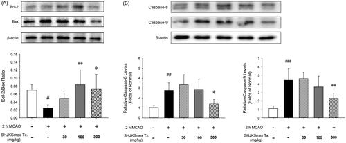
Figure 9. Representative photomicrographs of haematoxylin and eosin (H&E) staining (A) and quantitative analysis of the cell densities (B) of the hippocampal CA1 region in the brains of mice with middle cerebral artery occlusion (MCAO). Microscopic images of the whole brain (upper column) and the hippocampal CA1 region (lower column) from each group. Damaged cells were sparsely arranged. White bars in the lower column represent scale bars that indicate a length of 100 µm. Quantitative analysis showed significant changes in the pyramidal cell densities in the CA1 region. ###p < 0.001 vs. normal group and **p < 0.01 vs. control group; n = 5 in each group.
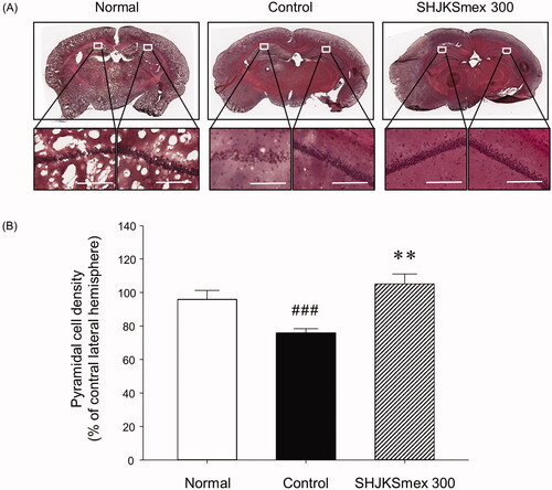
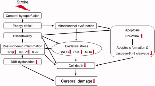
Figure 10. Schematic diagram of the antioxidant, anti-inflammatory, and anti-apoptotic mechanisms of the methanol fraction of the modified Seonghyangjeongki-san water extract (SHJKSmex) in mice with middle cerebral artery occlusion (MCAO). Red arrows represent the expected effects of SHJKSmex on the brain injury induced by MCAO.