
Figures & data
Figure 1. Effect of Rb1 on thoracic aorta senescence. Images of the immunohistochemical staining (400 ×) of p21Cip1 (A) and p16INK4a (B) in mouse thoracic aorta cross sections. (C) Western blot analysis of mouse thoracic aortic p21Cip1 and p16INK4a expression. The data are expressed as the mean ± SD. **p < 0.01 vs. the Young group; #p < 0.05 vs. the Old + Vehicle group.
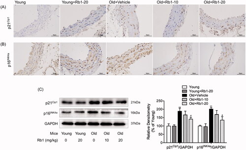
Figure 2. Rb1 treatment attenuated age-associated endothelium-dependent vasorelaxation impairment in thoracic aorta vascular rings. Figures showing the analysis results. The data are expressed as the mean ± SD. *p < 0.05, **p < 0.01 vs. the Young group; #p < 0.05, ##p < 0.01 vs. the Old + Vehicle group.
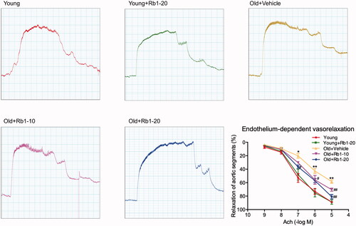
Figure 3. Rb1 reduced cellular adhesion molecule expression in aged mouse thoracic aortas. Western blot analysis of ICAM-1, VCAM-1 and PAI-1 expression in thoracic aorta tissues. The data are expressed as the mean ± SD. **p < 0.01 vs. the Young group; #p < 0.05, ##p < 0.01 vs. the Old + Vehicle group.
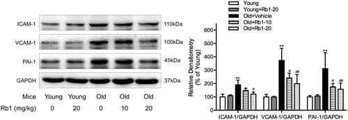
Figure 4. Rb1 treatment ameliorated ageing-induced vascular calcification and fibrosis. (A) Haematoxylin and eosin (HE) staining (400 ×), (B) Alizarin red S staining (400 ×) and (C) Masson’s trichrome staining (400 ×) of representative thoracic aorta sections in each group. (D) Western blot analysis of collagen I and collagen III expression in thoracic aorta tissues. The data are expressed as the mean ± SD. **p < 0.01 vs. the Young group; #p < 0.05, ##p < 0.01 vs. the Old + Vehicle group.
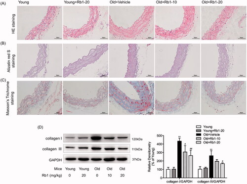
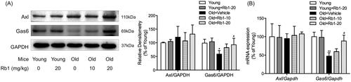
Figure 5. The influence of Rb1 on the Gas6/Axl signalling pathway. (A) Western blotting was performed to examine the protein expression of Gas6 and Axl. (B) qPCR was performed to examine the mRNA expression of Gas6 and Axl. The data are expressed as the mean ± SD. *p < 0.05, **p < 0.01 vs. the Young group; #p < 0.05 vs. the Old + Vehicle group.
Data availability statement
Data and materials are available upon request to the corresponding author.