Figures & data
Figure 1. 1,25-D3 promotes cell proliferation in H. pylori-infected GES-1 cells. (A) GES-1 cells were infected with H. pylori SS1 strain (MOI: 100) and treated with differernt concetrations of 1,25-D3 for 24 h, the cell viability was determined by CCK-8 assay. (B) GES-1 cells were infected with H. pylori SS1 strain (MOI: 100) and treated with different concentrations of 1,25-D3 for 24 h, LDH releasing was determined by a commercially available LDH assay kit. Bars represent means ± S.E.M of three independent experiments. ***p < 0.001 vs. H. pylori alone treatment.
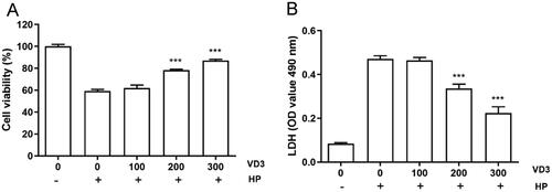
Figure 2. 1,25-D3 inhibits H. pylori-induced cell apoptosis in GES-1 cells. (A) GES-1 cells were infected with H. pylori SS1 strain (MOI: 100) and treated with different concentrations of 1,25-D3 for 24 h, the levels of apoptosis were analysed using an TUNEL detection kit. (B) GES-1 cells were infected with H. pylori SS1 strain (MOI: 100) and treated with different concentrations of 1,25-D3 for 24 h, caspase-3, caspase-6 and caspase-9 activities were determined by commercially available kits. (C) GES-1 cells were infected with H. pylori SS1 strain (MOI: 100) and treated with different concentrations of 1,25-D3 for 24 h, caspase-3, caspase-6 and caspase-9 expression were determined by western blot. Bars represent means ± S.E.M of three independent experiments. *p < 0.01 vs. H. pylori treatment.
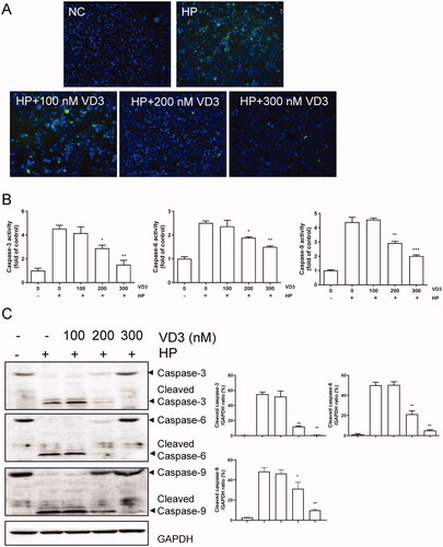
Figure 3. Bcl-2 families are involved in the anti-apoptotic effect of 1,25-D3 in H. pylori-treated GES-1cells. (A) GES-1 cells were infected with H. pylori SS1 strain (MOI: 100) and treated with different concentrations of 1,25-D3 for 24 h, Bcl-2, Bcl-xL, Bax and Bak levels were determined by western blot. (B) GES-1 cells were infected with H. pylori SS1 strain (MOI: 100) and treated with different concentrations of 1,25-D3 for 24 h, Cytochrome C (Cyto C) and apoptosis inducing factor (AIF) levels were determined by western blot. Bars represent means ± S.E.M of three independent experiments. *p < 0.01, **p < 0.05, ***p < 0.001 vs. H. pylori alone treatment.
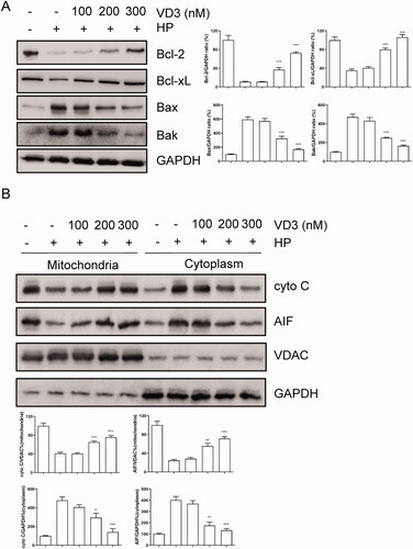
Figure 4. 1,25-D3 promotes c-Raf/MEK/ERK phosphorylation in H. pylori-treated GES-1 cells. GES-1 cells were infected with H. pylori SS1 strain (MOI: 100) and treated with different concentrations of 1,25-D3 for 24 h, (A) c-Raf, MEK and ERK phosphorylation levels and (B) RXRα and VDR levels were determined by western blot. Bars represent means ± S.E.M of three independent experiments. **p < 0.01, ***p < 0.001 vs. H. pylori alone treatment.
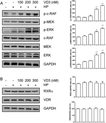
Figure 5. 1,25-D3 exerts an anti-apoptotic effect in H. pylori-treated GES-1 cells through binding to VDR. GES-1 cells were transfected with siRNA-VDR for 24 h, and then infected with H. pylori SS1 strain (MOI: 100) and treated with different concentrations of 1,25-D3 for 24 h, (A) the levels of VDR expression was determined by western blot, (B) the cell viability was determined by CCK-8 assay, (C) LDH release was determined by a commercial kit and (D) the levels of apoptosis were analysed using an TUNEL detection kit. Bars represent means ± S.E.M of three independent experiments. ***p < 0.001 vs. H. pylori treatment + siRNA-NC.
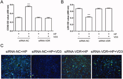
Figure 6. The inhibition of c-Raf/MEK/ERK phosphorylation blocks the anti-apoptotic effect of 1,25-D3 in H. pylori-treated GES-1 cells. GES-1 cells were treated with MEK inhibitor (U0126) for 24 h, and then infected with H. pylori SS1 strain (MOI: 100) and treated with different concentrations of 1,25-D3 for 24 h, (A) the cell viability was determined by CCK-8 assay, (B) LDH release was determined by a commercial kit and (C) the levels of apoptosis were analysed using an TUNEL detection kit. Bars represent means ± S.E.M of three independent experiments. ***p < 0.001 vs. H. pylori alone treatment, ###p < 0.001 vs. H. pylori + 1,25-D3 treatment.
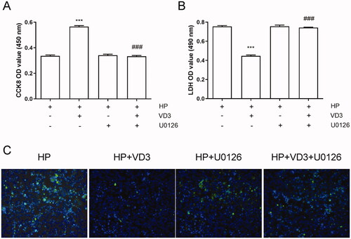
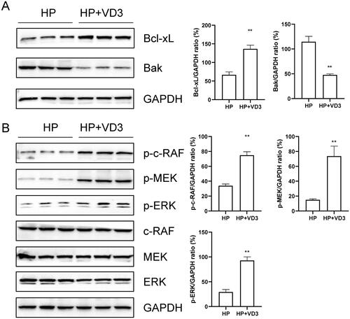
Figure 7. 1,25-D3 protects against H. pylori-infected apoptosis through a vitamin D receptor-dependent c-Raf/MEK/ERK pathway in mice. Mice were orally gavaged with 108 CFUs of H. pylori and 25 µg/kg 1,25-D3 every other day for 1 month. Bcl-xL, Bak, c-Raf, MEK and ERK phosphorylation levels in the stomach of mice were determined by western blot. Bars represent means ± S.E.M of three random mice. **p < 0.01 vs. H. pylori alone treatment.