Figures & data
Table 1. Primer sequences.
Figure 1. FA affects alveolar epithelial barrier function in sepsis-induced ALI. (A) Representative lung Section H&E staining images of mice that received sham operation or CLP. (B) Pathological score statistics of the three experimental groups. (C) Lung wet-to-dry ratios. (D) Total BALF protein concentrations measured by BCA assay. (E) MPO activity in lung tissues. (F) mRNA expression of tight junction proteins. (G) Protein expression of ZO-1, occludin, and claudin-1 in the lungs measured by western blotting. GAPDH served as an internal control. Original magnification 200×. *p < 0.05 (compared to the control group). #p < 0.05 (compared to the CLP group).
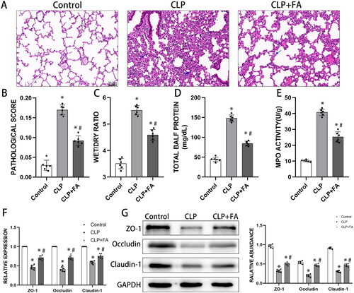
Figure 2. FA inhibits ferroptosis in sepsis-induced ALI. (A) Total iron levels in lung tissues. (B) MDA contents in different experimental groups. (C) GSH levels in lung tissues of different experimental groups. (D) mRNA expression of GPX4 measured with qPCR. (E) GPX4 protein expression was measured by western blotting, and GAPDH served as an internal control. *p < 0.05 (compared to the control group). #p < 0.05 (compared to the CLP group). $p < 0.05 (compared to the CLP + Fer-1 group).
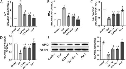
Figure 3. FA activates the Nrf2/HO-1 pathway in sepsis-induced ALI. (A) mRNA expression levels of Nrf2 and HO-1 in the lungs of CLP-treated mice were measured with qPCR. (B) Protein expression levels of Nrf2 and HO-1 in the lungs of CLP-induced mice were measured by western blotting, and GAPDH was used as an internal control. (C) The mRNA expression levels of Nrf2 and HO-1 in LPS-treated MLE-12 cells were measured with qPCR. (D) Protein expression of Nrf2 and HO-1 in LPS-treated MLE-12 cells was measured by western blotting, and GAPDH served as an internal control. *p < 0.05 (compared to the control group). #p < 0.05 (compared to the CLP or LPS group). $p < 0.05 (compared to the LPS + FA + sh-NC group).
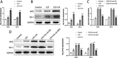
Figure 4. FA inhibits LPS-induced ferroptosis in alveolar epithelial cells by activating the Nrf2/HO-1 pathway. (A) Cell viability was determined with a CCK-8 assay. (B) Assessment of ROS levels in MLE-12 cells treated with LPS (500 ng/mL) for 24 h. (C) Detection of iron levels in MLE-12 cells treated with LPS (500 ng/mL) for 24 h. (D) MDA contents. (E) GSH levels. (F) mRNA expression of GPX4 measured with qPCR. (G) GPX4 protein expression was measured by western blotting, and GAPDH served as an internal control. *p < 0.05 (compared to the control group). #p < 0.05 (compared to the LPS group). $p < 0.05 (compared to the LPS + FA group). ⁁p < 0.05 (compared to the LPS + FA + sh-NC group).
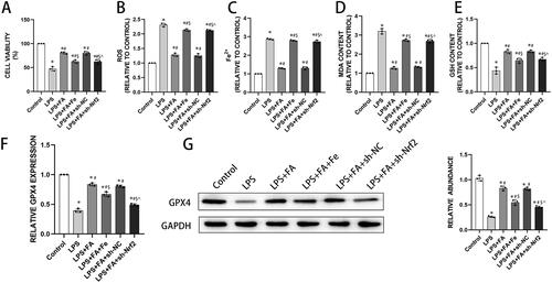
Figure 5. FA improves alveolar epithelial cell barrier function by repressing ferroptosis. Alveolar barrier function was measured with TEER (A) and FITC-dextran flux (B). (C) mRNA expression levels of ZO-1, occludin, and claudin-1 were measured with qPCR. (G) Protein expression levels of ZO-1, occludin, and claudin-1 were measured by western blotting, and GAPDH served as an internal control. * p < 0.05 (compared to the control group). #p < 0.05 (compared to the LPS group). $p < 0.05 (compared to the LPS + FA group). ⁁p < 0.05 (compared to the LPS + FA + sh-NC group).
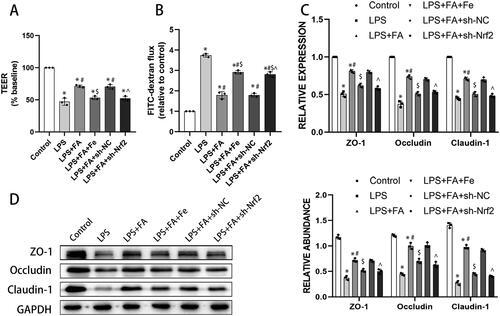
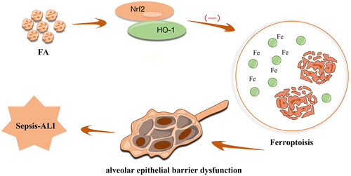
Figure 6. Schematic map of the role of FA in the modulation of sepsis-associated ALI. FA ameliorates ferroptosis-mediated alveolar epithelial cell barrier dysfunction by activating the Nrf2/HO-1 pathway.