
Figures & data
Figure 1. Representative photographs showing the (A) gross morphology, (B) absolute and (C) relative weights of the mouse seminal vesicles, and (D) fluid as compared among control, CUMS, and co-treated groups after co-treatment for 57 consecutive days (n = 12, each group). #p < 0.05, statistically significant difference as compared between the control and CUMS groups. *p < 0.05, statistically significant difference as compared between the CUMS and co-treated groups.
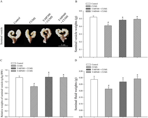
Figure 2. Representative microphotographs showing (A) histology of the seminal tissue and fluid strained by H&E, compared among control, CUMS and co-treated groups (T-MP 300 and 600 mg/kg BW). (Ba) normal seminal epithelium and histopathological features found in seminal vesicle of CUMS groups. (Bb) Decreasing of glandular epithelial cells. (Bc) Green arrows; pyknotic nuclei of basal cells, red asterisks; vacuolization within basal cells, blue asterisks; reduction of luminal seminal fluid.
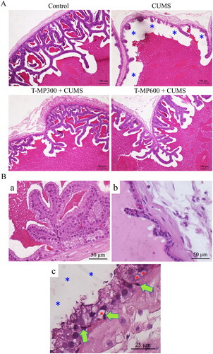
Figure 3. Immunofluorescence staining of the seminal vesicles compared among (Ab–c) control, (Ae–f) CUMS, (Ah–i) T-MP300 + CUMS, and (Ak–l) T-MP600 + CUMS groups against anti-androgen receptor (AR). Hoechst 33342 emitting blue fluorescence used as nuclear counterstain. AR immunostaining (green fluorescence). Expressions of (B) AR, heat shock protein 70 (Hsp70), and nuclear factor erythroid 2-related factor 2 (Nrf2) in seminal tissue compared among groups (n = 8, each group).
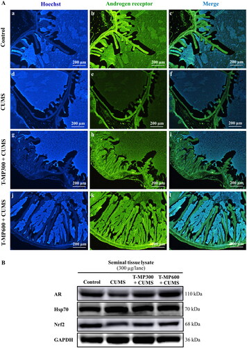
Figure 4. Expression of tyrosine phosphorylated (TyrPho) proteins observed in seminal tissue (A) and plasma lysates (B) compared among groups (n = 8, each group). EGF, epidermal growth factor used as positive control; BSA, bovine serum albumin used as negative control.
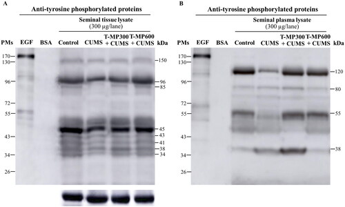
Figure 5. Immunofluorescence staining of seminal vesicle against anti-caspase 3 (A). Nuclei stained by Hoechst 33342 (blue-emitting fluorescent). Expression of pro and cleaved caspases 3 and 9 in rat seminal tissue and plasma lysates compared among control, CUMS and treated groups (B) (n = 8, each group).
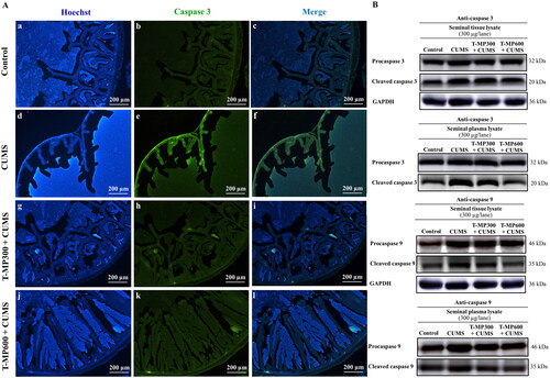
Table 1. Comparison of biochemical component parameters in the seminal fluid of the control, CUMS and co-treated groups (n = 8, each group).